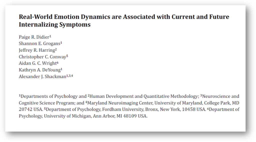


Shannon will soon depart us to take up her new post as Clinical Intern at Ohio State University in Columbus, OH. A bittersweet moment for a very proud academic family!
Kudos to graduate student, Shannon Grogans, who received the 2025 Michael J. Pelczar Award for Excellence in Graduate Study from the UMD Graduate School. The Pelczar Award is awarded to a doctoral candidate who has served at least one academic year as a teaching assistant with commendable performance, and who has demonstrated excellence beyond his or her course work.
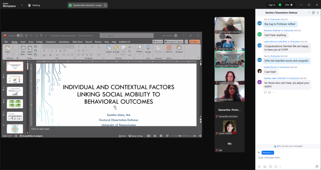
A hearty congratulations to our former postbac, Samiha Islam, on successfully defending her 3-study dissertation at the University of Pennsylvania under the mentorship of Sara Jaffee, and on learning that she was accepted into the clinical internship program at the Children’s Hospital of Philadelphia (CHOP). Mazel tov!!


Congrats to Shannon Grogans, graduate student recipient of the Department of Psychology’s 2024 Open Science Impactor Award. Shannon is being recognized for a collection of open science activities that she undertook for her Master’s thesis, which is currently under review at the Journal of Neuroscience. The list of activities is truly impressive and include, preregistration, sharing of unthresholded statistical maps on NeuroVault (a publicly accessible repository), posting of ROI level data on OSF, and continual updating of her preprint on bioRxiv. In addition, Shannon has been careful to document and share all deviations from her pre-registration plan, ensuring transparent and complete reporting of her results.
Congrats to Dr. Melissa Stockbridge, an alum of the lab, who recently secured her first faculty position in the Department of Neurology at Johns Hopkins University School of Medicine.
Assistant Professor | Neurology, Cerebrovascular Division
School of Medicine
Johns Hopkins University

| Welcome aboard to our incoming Ph.D. student, Kalina Kalinova! Kalina graduated from Sofia University in 2019 with an undergraduate degree in Psychology. After graduation, she completed her Masters degree in Clinical Neuropsychology at Leiden University and a research internship at the University of Cambridge, working with Dr. Kai Ruggeri. CV |
We are hiring a full-time postbaccalaureate research assistant (RA) to support our on-going NIMH and NIAAA studies. Read all about the position and living in the DMV here.
Updated April 30, 2024 — :We are currently reviewing applications and scheduling interviews. We are not accepting any additional applications at this time.
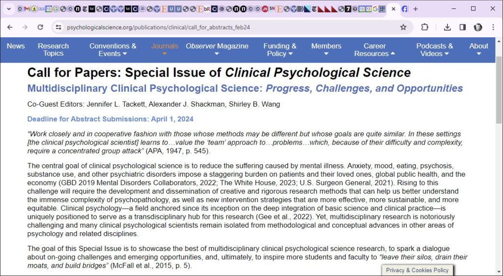
Like so many other scholarly, scientific, and medical disciplines, clinical psychology is often insular and inward facing, siloed off from the tools and conceptual frameworks of other scientific communities. Dr. Shackman is thrilled to serve as Guest Co-Editor of a special issue of Clinical Psychological Science focused on the challenges and opportunities of this often vexing, but rewarding endeavor. The Co-Editors need your help, in the form of illustrative empirical reports as well as more conceptual mini-reviews and opinion pieces. You need not be a bona fide clinical psychologist to participate! Psychiatrists, experimental psychopathologists, and other edge cases are welcome! Learn more at https://www.psychologicalscience.org/…/call_for…
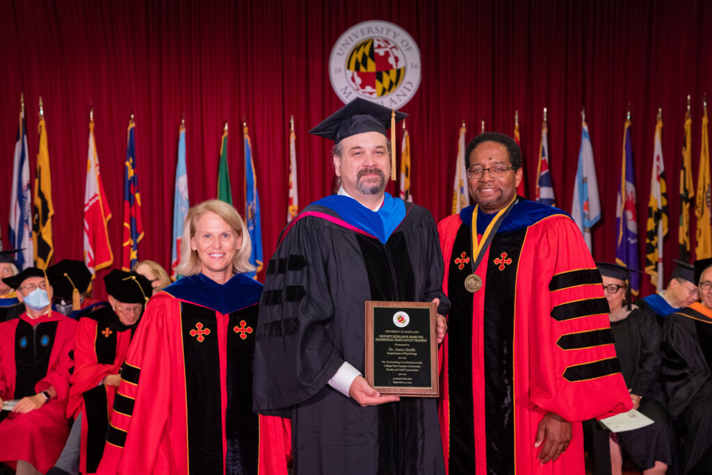
Congratulations to Dr. Jason Smith, recipient of the 2023 Provost’s Excellence Award for Professional Track Faculty. The award honor’s Dr. Smith’s many behind-the-scenes contributions to the University of Maryland neuroimaging community, to the Shackman and Blanchard labs, and to many other faculty and trainees at Maryland and other institutions in the US and abroad.

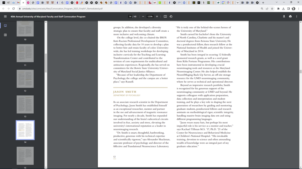
Read all about it here
Join us for a fun- and fact-filled Sunday morning continuing education (CE) event organized by the The Association of Practicing Psychologists: Montgomery – Prince George’s Counties and featuring none other than our very own, Dr. Shackman. Learn more about the event here.
A huge congrats to Shannon Grogans, who received a very fundable 1st percentile score on her NIMH Ruth L. Kirschstein Predoctoral Individual National Research Service Award application and was awarded the University of Maryland’s Ann G. Wylie Fellowship. Both awards will help support the final stages of her doctoral training and research, which are focused on understanding the relevance of fear- and anxiety-related brain circuits to the emergence and recurrence of pathological anxiety, depressed mood, and other internalizing symptoms in early adulthood. [05-25-2023 Update: Shannon’s F31 was funded and will begin in August 2023! She was compelled to decline the Wylie as a consequence.]
The lab was awarded a 5-year, $3M award from the NIAAA.
Alcohol misuse is a leading cause of human misery, morbidity, and mortality. Existing treatments are far from curative. While the roots of alcohol misuse are complex and multifactorial, Anxiety plays a key role. Models of addiction suggest that many drinkers misuse alcohol to relieve excess anxiety (‘self-medicate’). Yet the factors governing anxiety-fueled alcohol misuse remain incompletely understood.
This project will use a combination of techniques—including functional neuroimaging, computational modeling, and smartphone digital phenotyping—in a racially diverse sample drawn from the surrounding community to
In sum, alcohol use disorder and risky drinking, more generally, are notoriously heterogeneous, with thousands of unique clinical presentations. By focusing on a theoretically coherent set of dimensional measures in a diverse, clinically relevant sample, this project has the potential to overcome this barrier and provide fresh insights into the neurocomputational factors governing anxiety-fueled alcohol misuse. Building on well-established neuroeconomic models and a fruitful line of psychophysiological research, this study will provide a potentially transformative opportunity to identify new treatment targets; guide the development of new translational models; and inform the development of new digital interventions.
This project represents a collaboration between the Shackman laboratory and investigators at the University of Wisconsin—Madison (Dr. John Curtin) and University of California, Davis (Drs. Drew Fox and Erie Boorman).
We’re hiring! Learn more about the postdoc position here.

The lab was fortunate enough to receive a very generous new NIMH award focused on understanding the computational and neural architecture of fear and anxiety.
Despite growing concerns about validity, the NIMH Research Domain Criteria (RDoC) framework plays a key role in organizing basic, translational, and clinical research. RDoC’s approach to fear and anxiety is categorical: threat is either acute or potential; engages either the Amygdala or the bed nucleus of the stria terminalis (BST); and elicits either fear or anxiety.
Recent work by our team, Dean Mobbs, and other investigators casts doubt on this binary perspective, spurring the development of alternative approaches.
Dimensional models posit that threat responses vary along a smooth continuum of perceived danger—from absolutely safety to on-going attack. Danger perceptions are thought to emerge from parametric estimates of threat proximity, probability, and certainty, which are computed in weakly segregated cortico-subcortical circuits. To date, there have been no systematic, well-powered efforts to computationally implement these competing models and compare their validity.
Furthermore, while both models highlight the importance of threat uncertainty, they do not specify which kind. Computational psychiatry recognizes 2 mathematically distinct kinds of uncertainty: Risk and Ambiguity. Which of these is more relevant to threat reactivity and how they map onto the underlying neurobiology is unknown. To address these fundamental questions, we will recruit a racially diverse community sample enriched for elevated fear/anxiety symptoms. Using techniques adapted from neuroeconomics, a parametric threat-anticipation paradigm will allow us to simultaneously probe circuits sensitive to categorical (RDoC) and dimensional variation in threat for the first time. Smartphone phenotyping will assess real-world threat exposure, uncertainty, and distress.
Extreme fear and anxiety are leading causes of human misery and morbidity. This project will provide an exciting opportunity to develop one of the first computationally grounded models of fear and anxiety in a relatively large and diverse “DMV” (DC, MD, & VA) sample. It will help adjudicate on-going theoretical debates, validate a new conceptual approach for use with other read-outs and species, set the stage for new kinds of translational models and clinical studies, prioritize new targets for neuromodulation and other therapeutics development, and set the stage for the development of RDoC 2.0.
This project represents a team-science collaboration between the University of Maryland (Drs. Alex Shackman & Jason Smith) and the University of California-Davis (Drs. Andrew Fox & Erie Boorman)
You can read more about the project at Maryland Today.
Our NIMH-sponsored project focused on understanding the nature and neurobiology of paranoia was recently featured in a piece at Maryland Today. You can learn more about the project here.
We’re hiring! Learn more about the Study Coordinator position here.
A hearty congratulations to Shannon Grogans, who successfully completed her qualifying examination (“TIE Project”) and is now poised to propose her dissertation. Well done, Shannon!
Congratulations to Dr. Jason Smith on his well-deserved promotion to Associate Research Professor! Jason is truly exceptional and genuinely deserving of this promotion. He is a wonderful scientific partner and has made many important contributions to the Department of Psychology and the larger University of Maryland (UMD) neuroimaging community.
Jason has served as Director of Imaging Science in the Shackman Lab since 2014. In that time, he has played a leadership role, spearheading multiple research projects and grants, and providing thoughtful mentorship and training to students and faculty. Jason is hardworking, productive, creative, generous with his technical expertise, and scientifically rigorous. He is always willing to pitch in, whether that be volunteering to drive from his home in Bethesda to College Park for a crucial scanning session, submitting to multiple rounds of COVID testing to keep a sponsored project running through a global pandemic, or hosting a lab holiday party at his home.
Bringing Home the Bacon. At the time of his tenure review, Jason had served as a Co-Investigator, staff scientist, or consultant on millions of dollars of sponsored research projects led by UMD faculty (Blanchard, Dougherty, Hamilton, and Shackman), including 5 current NIH R01 awards and 4 prior awards. He also served as a Co-Investigator on 5 other R01 applications that were ultimately not funded.
An Inventive Scientist. Rachael Tillman’s master’s thesis is a classic example of Jason’s scientific ingenuity and support for clinical students (Tillman et al HBM 2018). The project leveraged publicly available fMRI data collected by Mike Milham’s group at the Nathan Kline Institute (Rockland sample). These data posed a number of unexpected technical challenges (e.g. how to optimally process idiosyncratically “de-faced” T1w anatomical scans), and Jason devised thoughtful solutions to each. Indeed, this project motivated him to further refine and optimize the MRI data processing pipeline used by my lab. Variants of Jason’s best-practices pipeline have since been adopted by a number of UMD labs, and Jason’s informal technical guidance influenced the development of fmriprep, an increasingly popular canned pipeline for fMRI data processing and quality control. He also recently organized a technical workshop, focused on quality control for fMRI, at the Czech National Institute of Mental Health.
Service to the UMD Neuroimaging Community and Beyond. Jason played a central role in the installation of the eye tracker and the electrical stimulator (‘shocker’) used by multiple groups at the Maryland Neuroimaging Center (MNC). He has since provided guidance on installation, best practices, and usage to investigators at UMD, UC Davis, Yonsei University, and elsewhere. Working closely with the MNC, Jason also contributed to early testing of advanced ‘multiband’ MRI pulse sequences at the MNC, sequences that are now routinely used by most UMD imaging labs. In addition, he currently serves as the system administrator and operational manager of the NeuroImaging Back-Up Server (NIMBUS), a secure off-site storage resource that services a number of UMD investigators (Bernat, Blanchard, Fox, Gard, Hamilton, Redcay, Riggins, and Shackman). Jason played a central role in establishing NIMBUS, which represents a collaborative partnership between the Shackman Lab, the Division of Information Technology (DIT), and the Office of Academic Computing Services (OACS). This involved innumerable emails and meetings with all of the various stakeholders. NIMBUS has allowed campus investigators to systematically back-up their imaging data, in some cases for the first time, while avoiding the high fees associated with alternative approaches, saving them tens of thousands of dollars.
Training & Mentorship. Jason is an outstanding trainer and mentor, and has played a crucial unsung role in numerous student projects, including 3 dissertations (McCarthy, Bradshaw, and Tillman), 3 master’s theses (Kaplan, Tillman, and Grogans), a NACS First Year Project (Kim), 2 senior theses, and multiple fellowship applications and awards (e.g. NSF GRF, NSF Combine, Wylie Dissertation Award). Jason has provided informal guidance and mentorship to graduate students in other labs and departments; to junior faculty, as they set up new imaging labs; and to senior faculty, as they embark on their first neuroimaging projects. He has also personally trained a half dozen full-time postbaccalaureate research assistants and dozens of undergraduate research assistants.
Conclusion. In sum, Dr. Jason Smith is an outstanding Research Scientist and has played a crucial role in our lab’s success and that of many other faculty and trainees on and off campus. He’s an exceptional contributor and one of the behind-the-scenes heroes of the college. Congrats on this well-deserved recognition!
Please join us for several upcoming presentations…

Congratulations to Shannon Grogans, who successfully proposed her TIE (Transition to Independence and Expertise) project. The TIE project is a key milestone in the clinical psychological science doctoral program at Maryland. The TIE project, which replaces the more traditional qualifying exams, serves as the gateway to the dissertation. For her TIE project, Shannon will prepare a NIH NRSA (“F31”) fellowship application focused on linking neuroimaging measures of threat-reactivity to longitudinal waves of clinical, life stress, and smartphone experience-sampling (‘EMA’) data in young adults at risk for developing anxiety disorders and depression. As part of the TIE experience, Shannon will receive structured mentorship in grant preparation from several faculty (Dougherty and Chronis-Tuscano) with successful records of NIH support. If awarded, the NRSA will provide protected time, allowing Shannon to more fully immerse herself in advanced statistical modeling and EMA techniques. Independent of the ultimate outcome of the fellowship application, the TIE project will allow her to develop a detailed blueprint for her dissertation project.
A hearty congratulations to Gloria Kim, who passed the NACS qualifying exams, opening the door to proposing her doctoral dissertation project. She’s now ABD! The interdisciplinary exams were co-chaired by Gloria’s co-supervisors, Drs. Arianna Gard and Alex Shackman.
Shannon’s pre-registered study was chosen as the winner of the first annual Reproducibility award at the annual meeting of the Society for Social Neuroscience (S4SN). Congrats, Shannon!
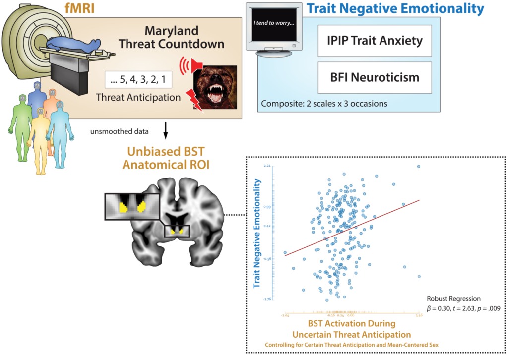
The central focus of the project was on clarifying the neural circuits underlying individual differences in Neuroticism/Negative Emotionality (N/NE). N/NE — the tendency to experience and express more intense, frequent, or persistent negative emotions — is a central dimension of mammalian temperament, and elevated levels of N/NE confer risk for a wide variety of adverse outcomes, from dissatisfaction and divorce to disease and premature death. The deleterious consequences of N/NE are particularly evident in the sphere of mental illness. Individuals with a more negative disposition are more likely to develop anxiety disorders and depression and, if they do, to experience more severe, chronic, and intractable symptoms. Yet the neural systems underlying trait-like individual differences in N/NE have remained enigmatic, impeding the development of more effective or tolerable interventions.
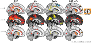
Shannon used a suite of neuroimaging and data science techniques to test the hypothesis that N/NE reflects heightened reactivity to impending threat in the central extended amygdala, including the central nucleus of the amygdala (Ce) and bed nucleus of the stria terminalis (BST). To ensure a broad spectrum of N/NE, 220 young adults were selectively recruited from a pool of nearly 7,000 pre-screened individuals. A multiband pulse sequence, best-practice data processing techniques, spatially unsmoothed data, anatomically defined regions-of-interest, and a cross-validated robust-regression framework enabled unbiased hypothesis testing with unprecedented anatomical resolution.
To clarify specificity, parallel analyses were performed using data from an emotional-faces paradigm. While ‘threatening’ faces are potent triggers of amygdala activity and widely used in on-going biobank projects, they do not elicit robust negative affect and are better conceptualized as a probe of emotion perception.
Results revealed that N/NE is associated with heightened BST activation during the anticipation of aversive stimulation, and this association is uniquely evident when the timing of aversive stimulation is uncertain. BST reactivity to uncertain threat remained predictive of N/NE after controlling for BST reactivity to certain threat or Ce reactivity to uncertain threat. In contrast, N/NE was unrelated to Ce activation during threat anticipation and BST/Ce reactivity to emotional faces.
While it is tempting to treat different ‘threat’ paradigms—from viewing photographs of ‘threatening’ faces to anticipating the delivery of noxious stimulation—as interchangeable probes of brain function, this has rarely been examined.
Follow-up analyses revealed no evidence of between-task convergence in the amygdala (Ce) and marginal evidence in the BST.
A large, sample and pre-registered, best-practices approach enhance confidence in the robustness and translational relevance of these results.
Together, these observations have implications for theory and practice and lay the groundwork for the longitudinal and mechanistic studies that will be necessary to determine causation and, ultimately, to develop improved interventions for extreme N/NE.
Grogans, S. E., Hur, J., Barstead, M. G., Anderson, A. S., Islam, S., Kim, H. C., Kuhn, M., Tillman, R. M., Fox, A. S., Smith, J. F., DeYoung, K. A., & Shackman, A. J. (2021). Neuroticism/negative emotionality is associated with increased reactivity to uncertain threat in the bed nucleus of the stria terminalis. Poster presented at the annual meeting of the Society for Social Neuroscience.
Congratulations to Shannon Grogans, who successfully defended her (pre-registered) Master’s thesis, entitled “Understanding the Relevance of Extended Amygdala Reactivity to Dispositional Negativity.”
As Shannon notes in the thesis, dispositional negativity—the propensity to experience and express more intense, frequent, or persistent negative affect—is a fundamental dimension of mammalian temperament. Elevated levels of dispositional negativity are associated with a wide range of practically important outcomes, from marital stability and socioeconomic attainment to anxiety disorders and depression. Yet our understanding of the brain bases of dispositional negativity remains far from complete. Converging lines of mechanistic and neuroimaging evidence suggest that dispositional negativity reflects heightened threat reactivity in the extended amygdala—a circuit encompassing the dorsal amygdala in the region of the central nucleus (Ce) and the neighboring bed nucleus of the stria terminalis (BST)—and that this association may be more evident when threat is uncertain.
To address this, Shannon used a combination of approaches—including a multi-trait, multi-occasion composite of dispositional negativity and neuroimaging assays of threat anticipation and perception—to demonstrate that individuals with a more negative disposition show heightened BST activation during threat anticipation. A series of cross-validated, robust regression analyses revealed that dispositional negativity is uniquely predicted by BST reactivity to uncertain-threat anticipation. In contrast, dispositional negativity was unrelated to amygdala activation during the anticipation of threat or to extended amygdala activation during the presentation of ‘threatening’ faces.
As Shannon notes, these brain imaging observations lay the foundation for the kinds of prospective-longitudinal and mechanistic studies that will be necessary to determine causation and, ultimately, to develop improved interventions for extreme dispositional negativity.
Open Science FTW! Although the lab has long made a point of uploading raw data to NDA for select NIMH-sponsored work and 2nd-level fMRI stats maps to NeuroVault for all publications, we are now taking it to the next level, and beginning to share subject-level brain imaging data with the ENIGMA Anxiety Workgroup (ENIGMA-Anxiety) and with the Affective Neuroimaging Collaboratory. These data will ultimately enable well-powered, reproducible, multi-lab analyses of the brain bases of anxiety disorders and BI, and the development of more sensitive and specific brain signatures of emotion. This is important because emotional disorders impose a staggering burden on global public health, and existing interventions are inconsistently effective.
Data sharing is a core scientific value, and maximizes the return on precious taxpayer investments in basic and clinical research.
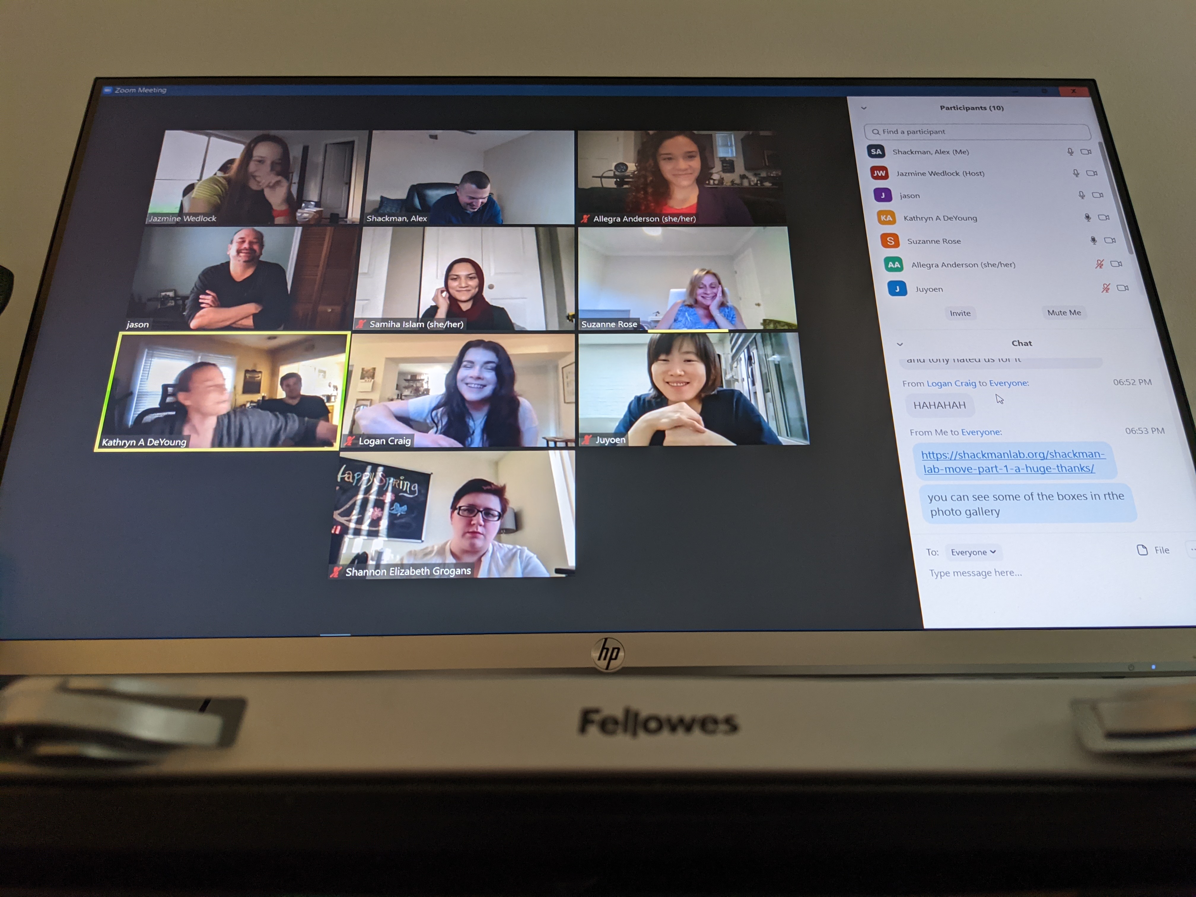
Katie wrote, It’s crazy to think that it’s been almost a full year since the COVID-19 pandemic shut down the world as we knew it. Despite the extreme uncertainty that we’ve all faced over the last year, we’ve continued to persevere and put our best foot forward. In recognition of this, I wanted to share an exciting announcement of what we have accomplished.
As of March 2021, we have finished data collection for our two and a half year longitudinal study entitled “Prospective determination of neurobehavioral risk for the development of emotion disorders,” also affectionately known as “The neuroRED Project” or “PAX.”
Beginning in the Fall of 2016, our team set out to better understand how emotions are organized in the brain, how they differ from person to person, and ways in which these differences can influence the risk of developing an anxiety disorder or depression. Our recruitment and baseline data collection efforts included the screening of over 6,000 prospective participants, 258 consents, 246 lab assessments and smartphone ecological momentary assessment (EMA) batteries, 250 clinical assessments, and 241 MRI sessions. The volume and speed at which fMRI data collection was conducted was so extraordinary that the Maryland Neuroimaging Center (MNC) called for a reform of MRI scheduling procedures. These efforts ultimately resulted in 234 participants who were eligible for the longitudinal study.
Since then, we have completed 4 follow-up data collections, including 690 lab assessments, 691 EMA batteries, and 460 clinical assessments. In the end, we achieved an overall retention rate of 97.86% – an amazing feat for a longitudinal study (see table below for more details). Our participants enjoyed being a part of this project to the extent that 96.58% of them reported that they would like to be contacted for future research opportunities with the Shackman lab. Further, this study has provided our students with a number of invaluable training opportunities, from learning how to collect and make sense of cutting-edge brain imaging data to public speaking.
All of this is an absolutely enormous accomplishment that could not have been done without the help of each and every one of you. Through tight deadlines, long weekends at the MNC, countless hours spent on the phone, and hundreds of emails… we did it! As we like to say to our participants: “Thank you so much for your continued participation in the neuroRED project over the past two-and-a-half years. Your contributions were critical to our success and have already begun to provide important new insights to the fields of psychology and neuroscience research!”
But seriously… I hope you can join us in feeling the pride, joy, and relief that we are feeling right now. This has been a remarkably rewarding journey for all of us in the lab – scientifically, professionally, and personally. Alex and I are eternally grateful for your willingness to share so much of yourselves and your time with us.
Alex wrote, First, a deep bow of gratitude to each and every one of you for making this possible. I would like to extend a special, heartfelt thanks to
-> Sue Rose, who patiently helped guide the many revisions we made to the standard SCID-5 modules, single-handedly completed hundreds of detailed clinical interviews, and was compelled to repeatedly drive to the UPS store lugging the resulting boxes of interview notes
-> Jason Smith, who built the imaging paradigms and devised new ways of processing, analyzing, and storing the data
-> Katie DeYoung, the heart and soul of the project. Katie was involved with every aspect of PAX, often working extraordinarily long hours to ensure its success. Paired with Sue’s gentle probing of subjects’ psychological innards, Katie’s cheerful case-management approach made it possible to maintain extraordinarily high levels of sample retention across multiple waves of assessment.
You three made it possible for me to maintain my sanity and (for the most part) sleep soundly at night over the past 5 years, and we and other researchers will reap the scholarly rewards of your extraordinary efforts for years to come. I am so very grateful.
Thanks to EVERYONE who contributed to this project, in ways small or large. We could not have done it without you.
The lab is moving! Just around a couple of corners to a larger suite of rooms in the Department of Psychology. A deep bow of gratitude to Jazmine Wedlock and Katie DeYoung for spearheading our egress, and to Mike Dougherty and Joanne Leffson-Bryant for helping with the administrative details.
Rachael Tillman — who currently is currently completing a clinical internship at Dell Children’s Medical Center in Austin, Texas — has accepted a 2-year post-doctoral fellowship in Pediatric Neuropsychology at Children’s National Hospital in DC. Congrats, Rachael!
Postdoctoral Position in Affective and Clinical/Translational Neuroscience
University of Maryland, College Park, MD
Candidates are being considered for a 1-year NIMH-funded postdoctoral position in the laboratory of Dr. Alex Shackman in the Department of Psychology at the University of Maryland at College Park (http://shackmanlab.org/). For U.S. citizens residing in North America, we are open to this being a largely remote (‘tele-work’) position.
The focus of this position will be to support on-going analytic and manuscript projects focused on the neurobiology of fear and anxiety and its role in the development and maintenance of anxiety disorders, depression, substance abuse, and psychosis. A secondary focus will be linking variation in circuit function to thoughts, feelings, and behavior in the clinic and real-world, indexed via semi-structured interviews and smartphone-based ecological momentary assessment (EMA). In most cases, data have already been collected. There will be opportunities to become involved in other projects, develop new analytic strategies, and develop mentored fellowship and grant applications (e.g. F32/K).
We are particularly excited about candidates with excellent writing, organizational, and statistical skills, and a strong background in functional neuroimaging and affective/clinical neuroscience. Expertise in HLM/MLM and related analytic techniques (e.g. SEM) or machine learning is desirable but not necessary. We want someone who is comfortable teaching themselves new techniques and who can jump right into doing science, so decent-or-better technical skills are mandatory. Applicants should have a Ph.D. in a relevant field and excellent organizational, interpersonal, and writing skills. This is a 1-year position that is potentially renewable, contingent on performance and funding.
Applicants should send a cover letter or portfolio describing relevant skills, experience, and interests—please provide concrete details about your technical contributions to past projects. Please include a current CV and contact information for 2-3 references to Dr. Shackman (shackman@umd.edu); the Director of Laboratory Operations, Kathryn DeYoung (deyoung.k@gmail.com); and the Laboratory Manager, Jazmine Wedlock (jwedlock@umd.edu). Applicants will be considered until the position is filled. The University of Maryland is an Equal Opportunity/Affirmative-Action Employer. Read more about the NIMH R01 project here and here. Read more about the NIDA R21 project here and here.
This is a fantastic opportunity to live in and explore DC, MD, and Northern VA!
The University of Maryland is an equal opportunity affirmative action employer with a commitment to racial, cultural, and gender diversity. We do not discriminate in hiring on the basis of sex, gender identity, sexual orientation, race, color, religious creed, national origin, physical or mental disability, protected Veteran status, or any other characteristic protected by federal, state, or local law.
A new study from the Shackman lab in the Journal of Neuroscience has been featured in several recent stories in the media. Building on other work by our group over the past decade (for example, this theoretical review), the new brain imaging study provides evidence that uncertain and certain threat are processed by a common core threat anticipation network. These results run counter to the popular idea that waiting for certain (“fear”) and uncertain threat (“anxiety”) reflect strictly segregated, non-overlapping brain circuits.
The study, which was first selected for special promotion by the Society for Neuroscience and UMD Brain & Behavior Institute, has since been the focus of articles in Axios, The Scientist, Medical Xpress, SciTech Daily, Theravive, Maryland Today, and the Italian-language Le Scienze.
The study was supported by the National Institute of Mental Health, and spearheaded by our former postdoc, Dr. Juyoen Hur (now an Assistant Professor at Yonsei University, Seoul, South Korea).
Interested in learning more? The paper is freely available here.
A hearty congrats to Gloria Kim, who successfully proposed her NACS First Year Project!
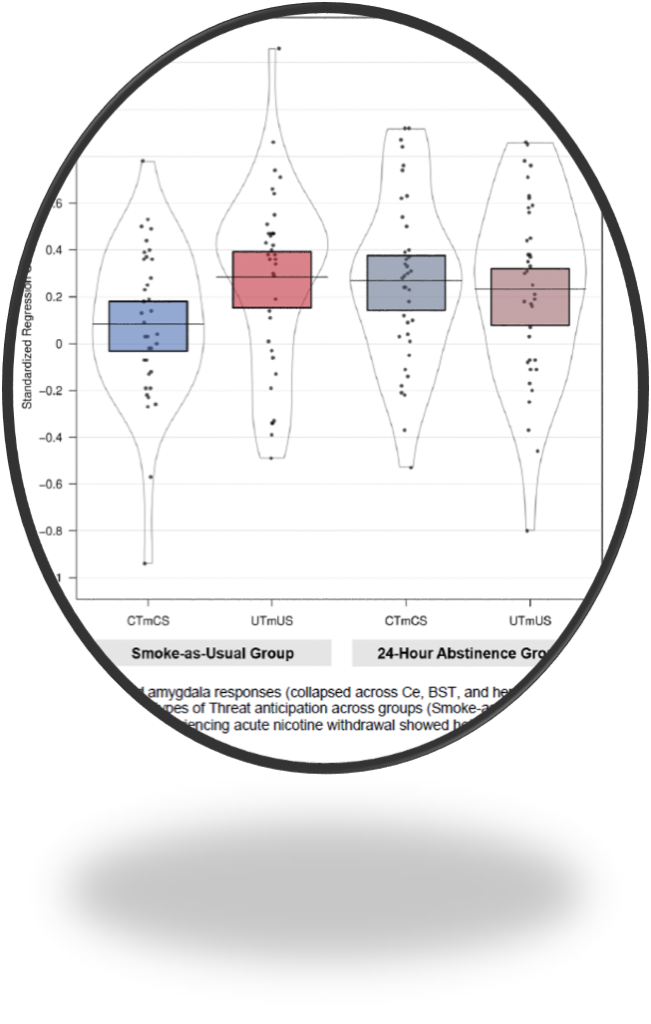
Gloria’s NIDA-sponsored project is focused on understanding the contributions of the extended amygdala to heightened negative emotion and amplified stress reactivity in tobacco smokers experiencing acute nicotine withdrawal during a period of voluntary abstinence.
A hearty congrats to Shannon Grogans, who successfully proposed her master’s thesis!
Shannon’s project is focused on understanding the neural systems underlying trait-like individual differences in dispositional negativity, a prominent risk factor for anxiety disorders, depression, and a host of other adverse outcomes. A secondary focus of her project is to determine whether different experimental probes of extended amygdala function are interchangeable. This is an important question because it is widely assumed that different ‘threat’ tasks— from viewing photographs of fearful faces to waiting to receive a painful electric shock—provide equivalent measures of amygdala reactivity, an assumption that has led large on-going biobank studies to adopt the more convenient faces approach. We wish her the best of luck as she begins this new chapter of her professional training.

A hearty congrats to Kathryn DeYoung, who submitted her Master’s thesis!
Katie’s project leveraged survey data collected from 187,000 respondents over an 11 year span to provide the most compelling evidence to date that graduate school confers heightened risk for emotional distress, emotional disorders, and suicidality. Her results also make it clear that the prevalence of these problems has increased over the past decade. Using data gleaned from the NSDUH survey, an annual randomized household interview study of mental illness, she rigorously demonstrates that American graduate students are more likely to experience depression and suicidality than their demographically matched peers, and that this discrepancy has worsened since 2008. In sum, her results paint a worrisome portrait of the state of graduate student mental health in the United States, with important implications for the full spectrum of stakeholders, from students and faculty to administrators and funders. We wish Kathryn the best of luck with the coming oral defense.
December , 2020 update: She passed! Thesis accepted with minimal revisions!

New research from the lab focused on understanding the neural circuits underlying fear and anxiety was featured in Smithsonian magazine.
Dr. Shackman was selected as a fellow of the Association for Psychological Science, the leading international organization dedicated to advancing basic and applied scientific psychology.
Paranoid ideation—the mistaken belief that intentional harm is likely to occur—spans a continuum, from mild suspicion to persecutory delusions. Among patients with schizophrenia and other psychosis disorders, elevated levels of paranoia are common, debilitating, and challenging to treat.
The cues (public environments, strangers) and processes (anxiety) that promote paranoia have grown increasingly clear, but the brain bases of these pathways are unknown, thwarting the development of mechanistic models and, ultimately, the development of more effective or tolerable biological interventions.
This multi-disciplinary project will use a combination of tools to clarify the factors governing paranoia. We will enroll the full spectrum of paranoia without gaps or discontinuities—including psychosis patients with frank persecutory delusions and matched community controls. These data will allow us to rigorously examine the hypothesized contribution of brain circuits responsible for triggering anxiety and evaluating the threat potential of everyday social cues. Integrating neuroimaging measures with smartphone experience-sampling data will enable us to extend these insights to the real world for the first time. It has become increasingly clear that traditional psychiatric diagnoses, like Schizophrenia, present significant barriers to understanding etiology. Our focus on dimensional measures of paranoia overcomes many of these barriers and dovetails with the HiTOP framework.
This project is led by Drs. Jack Blanchard, Jason Smith, and Alex Shackman at Maryland, and encompasses a collaboration with Dr. Alan Anticevic‘s group at Yale. The award is sponsored by the National Institute of Mental Health.
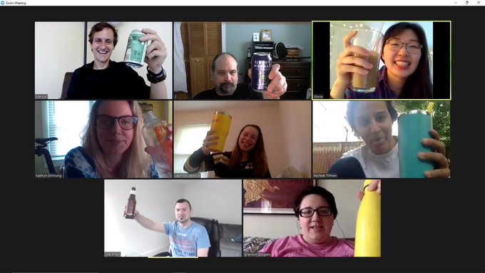
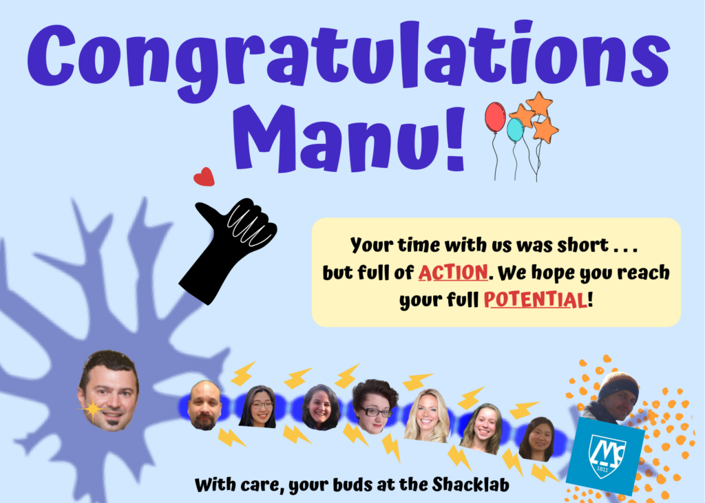
Bittersweet congrats to Manuel (‘Manu’) Kuhn! New postdoctoral position at McLean with the Pizzagalli group.
To quote, Dr. Smith (top-center), “Manuel, we’ve enjoyed having you here. You’ve been a pleasure to have in the lab. I know the graduate students have benefited immensely from having you here to help them. When we both sent code to extract an ROI to help an unnamed grad-student, they used yours because it “made more sense.” That is a valuable thing. Other things you’ve given to them have also “made sense.” There is something special about knowing you have left a mark on students as they move forward and I think you have left something here. It’s a nice thing to hold on to when you’re fighting with reviewers on your next paper. You’ve also helped me with looking back at the big picture. It’s easy (and fun) to get lost in complex solutions to at least seemingly simple problems. It’s worth it to be reminded that rather than always hunting more and more complex solutions, simple ones can also have utility. It was humbling to see your “let’s just do it” SPM/SPM results pop as expected when JFS/JFS results were null no matter what the ultimate reason and outcome. There is definitely something to be learned there and I will try to remember it. I hate being wrong with all of my being but I absolutely love it too and would never pass it up so, thank you. I hope the totality of the AAS experience we’ve had gives you that same perspective. Seeking knowledge is difficult. Mostly we fail miserably. Maybe, once in a blue moon, we hit something truly interesting. Even if it never sees publication, that new piece of knowledge you’ve found it worth all the effort.
That said, you BETTER get something together to submit … within the next month or I’m coming up to Boston with a big stick to hunt you down! We’ve enjoyed having you in the lab and I wish you all the best in the future.”
Shannon Grogans’ revised application was selected for an honorable mention (top 25%) by the National Science Foundation. This is an extremely competitive national award, with more than 12,000 applications submitted every year. Congratulations, Shannon!!!


A hearty congratulations to Dr. Juyoen Hur, who will be taking up her new post as an Assistant Professor in the Department of Psychology at Yonsei University in her hometown of Seoul, South Korea, just in time for the start of the Spring 2020 academic term. Yonsei is among the three ‘SKY’ universities, widely considered the most prestigious in Korea. While we will greatly miss Juyoen’s kindness and warmth, we are filled with joy, knowing that she will be so close to friends and family after a decade of training in the United States. We look forward to future collaborations, to crossing paths at scientific meetings, and to seeing her new laboratory flourish!

Gloria Kim, a first-year graduate student in the lab, was awarded a
Computation and Mathematics for Biological Networks (COMBINE) fellowship. COMBINE is the University of Maryland’s National Science Foundation-funded Research Traineeship (NRT) program in Network Biology. COMBINE immerses doctoral students in interdisciplinary research and training that integrates quantitative modeling methods from physics and mathematics with data processing, analysis, and visualization tools from computer science to gain deeper insights into the structural and dynamical principles governing living systems. Participants learn to utilize a network-based, data-driven approach, focusing on how interaction patterns can give insights into complex biological phenomena. COMBINE prepares students to become experts in the process of transforming raw biological data into useful information from which new biological insights can be inferred, positioning them to pursue a range of Science, Technology, Engineering, and Mathematics (STEM) careers at the nexus of the computer, physical, and life sciences. Fellows receive training in 4 areas of network analysis: (a) quantitative metrics for biological networks, (b) mechanistic models of biological networks, (c) network statistics and machine learning for biological applications, and (d) and visualization techniques for large, complex biological data sets. Dr. Rasmus Birn (Departments of Psychiatry & Medical Physics, University of Wisconsin, Madison) will serve as the out-of-field mentor for Gloria’s fellowship. Learn more about COMBINE.
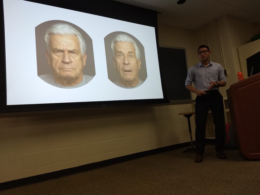
Richard, a longtime research assistant in the lab, successfully defended his senior thesis, “Dispositional negativity and the central extended amygdala.” Dr. Shackman chaired the thesis committee, which included Drs. Jens Herberholz and Quentin Gaudry. The thesis was accepted without revision and received the honors designation to boot. Congrats, Richard! We’re proud of you!
The University of Maryland has awarded Rachael Tillman a prestigious Wylie Dissertation Fellowship for the 2019-2020 academic year. Congrats, Rachael!
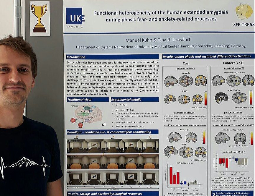
Dr. Manuel Kuhn will be joining our team in July 2019! Welcome aboard, Manuel!
This is a fantastic opportunity to live in and explore DC, MD, and Northern VA!
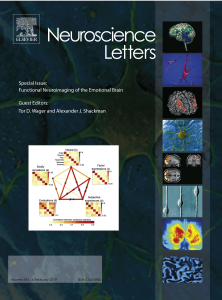 Emotions are central to who we are. They influence our worldviews, decisions, and memories and play a key role in determining which of the myriad events bombarding our senses we see, hear, and feel. Emotions arise in direct proportion to personal meaning—how relevant a stimulus or situation is to our well-being. They are thought to be hallmarks of integrative brain processes that bind together perceptions of the world, interoception of our bodily states, conceptions of meaning, and prospections about the future. Their integrative nature also makes them complex, and for decades the brain processes that underlie emotions have remained part of a dark unknown space in our understanding of the mind. However, recent years have witnessed a resurgence of interest in the organization of emotional brain systems. Numerous papers, reviews, and books—academic and popular-press—deconstruct the ‘emotional brain’ and its implications for health, disease, and other kinds of practically important outcomes. Our Special Issue of Neuroscience Letters, Functional Neuroimaging of the Emotional Brain (Guest Edited by Tor Wager & Alex Shackman), surveys recent advances in this burgeoning area of research, with leading investigators from across North America and Europe contributing 10 original theoretical reviews. This exciting body of work encompasses a broad spectrum of populations—from rodents and monkeys, to children and psychiatric patients—and showcases a wide variety of paradigms, measures, analytic strategies, and conceptual approaches.
Postdoctoral fellow, Juyoen Hur, was accepted into the 2019 ADAA Career Development Leadership Program. Congrats, Juyoen!
Dr. Shackman is accepting graduate student applications this cycle. Although you are welcome to apply via the Clinical Psychology area group in the Department of Psychology or the Neuroscience and Cognitive Science (NACS) training program, we strongly prefer NACS (‘affective neuroscience’) applications. This is a great opportunity to live in and explore the greater DC, MD, Northern VA region (‘The DMV’)!
Here are some tips–good luck!
Letters to potential advisors: Steve Luck’s Blog (I will respond slowly, but I promise that I will read and respond!)
Reflections on Graduate School: Blog. (Most of what Russ writes here applies equally well to our lab!)
PhD Life:
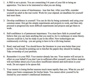
You might also check out this inspirational piece by Kay Tye (MIT): Mission
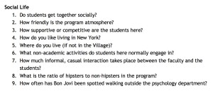
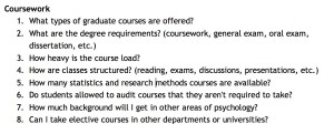
Some Questions to Ask Potential Mentors (h/t Jay Van Bavel, NYU):

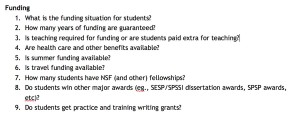
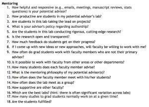
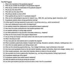
What have researchers learned about the roots of human emotion? That’s the focus of the recently published “The Nature of Emotion”. Co-editor Alex Shackman shares the latest research on the mind and our understanding of our emotions in this interview.
A new paper from the lab was selected for special promotion by the Journal of Neuroscience and has since been picked up by a number of local and national media outlets, including the University of Maryland, ScienceNews, Newsweek, and The Diamondback.
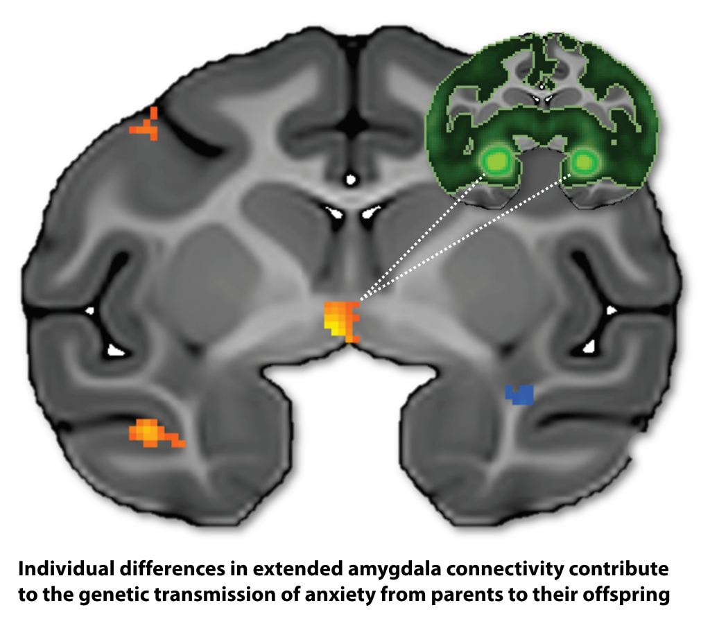
If you’re planning to attend the upcoming SRP meeting in Indianapolis, please consider extending your visit to the heartland by 36 hours and participating in the HiTOP 2018 satellite meeting on Sunday and Monday.
The satellite meeting will be on September 23rd-24th, 2018. On Sunday the 23rd we will meet in the afternoon and on Monday the meeting will be a full day of programming and work group activities.
To learn more about HiTOP and the urgent need for next-gen nosology, check out these new papers:
Krueger, R. F., Kotov, R., Watson, D., Forbes, M. K., Eaton, N. R., Ruggero, C. J., Simms, L. J., Widiger, T. A., Achenbach, T. M., Bach, B., Bagby, R. M., Bornovalova, M. A., Carpenter, W. T., Chmielewski, M., Cicero, D., Clark, L. A., Conway, C., DeClercq, B., DeYoung, C. G., Docherty, A. R., Drislane, L. E., First, M. B., Forbush, K. T., Hallquist, M., Haltigan, J. D., Hopwood, C. J., Ivanova, M. Y., Jonas, K. G., Latzman, R. D., Markon, K. E., Miller, J. D., Morey, L. C., Mullins-Sweatt, S. N., Ormel, J., Patalay, P., Patrick, C. J., Pincus, A. L., Regier, D. A., Reininghaus, U., Rescorla, L. A., Samuel, D. B., Sellbom, M., Shackman, A. J., Skodol, A., Slade, T., South, S. C., Sunderland, M., Tackett, J. L., Venables, N. C., Waldman, I. D., Waszczuk, M. A., Waugh, M. H., Wright, A. G. C., Zald, D. H. & Zimmerman, J. (2018). Progress in achieving empirical classification of psychopathology. World Psychiatry, 17, 282-293. PDF ** Focus of nine accompanying commentaries PDF
Conway, C. C., Forbes, M. K., Forbush, K. T., Fried, E. I., Hallquist, M. N., Kotov, R., Mullins-Sweatt, S. N., Shackman, A. J., Skodol, A. E., South, S. C., Sunderland, M., Waszczuk, M. A., Zald, D. H., Afzali, M. H., Bornovalova, M. A., Carragher, N., Docherty, A. R., Jonas, K. G., Krueger, R. F., Patalay, P., Pincus, A. L., Tackett, J. L., Reininghaus, U., Waldman, I. D., Wright, A. G. C., Zimmerman, J., Bach, B., Bagby, R. M., Chmielewski, M., Cicero, D. C., Clark, L. A., Dalgleish, T., DeYoung, C. G., Hopwood, C. J., Ivanova, M. Y., Latzman, R. D., Patrick, C. J., Ruggero, C. J., Samuel, D. B., Watson, D. & Eaton, N. R. (revision under review). A Hierarchical Taxonomy of Psychopathology can reform mental health research. Preprint available at psyarxiv.com PDF
Ruggero, C. J., Kotov, R., Hopwood, C., First, M., Clark, L. A., Skodol, A., Mullins-Sweatt, S. N., Patrick, C. J., Bach, B., Cicero, D., Docherty, A., Simms, L. J., Bagby, M., Krueger, R. F., Callahan, J., Chmielewski, M., Conway, C., DeClercq, B. J., Dornbach-Bender, A., Eaton, N., Forbes, M., Forbush, K., Haltigan, J. D., Miller, J. D., Morey, L. C., Patalay, P., Regier, D., Reninghaus, U., Shackman, A. J., Shteynberg, Y., Waszczuk, M. A., Watson, D., Wright, A. G. C., Zimmerman, J. (under review). Integrating the Hierarchical Taxonomy of Psychopathology into clinical practice. Preprint available at psyarxiv.com PDF
Waszczuk, M. A., Eaton, N. R., Krueger, R. F., Shackman, A. J., Waldman, I. D., Zald, D. H., Lahey, B. B., Patrick, C. J., Conway, C. C., Ormel, J., Hyman, S. F., Robinson, E. B., Fried, E. I., Forbes, M. K., Althoff, R. R., Bach, B., Chmielewski, M., DeYoung, C. G., Docherty, A., Forbush, K. T., Hallquist, M., Hopwood, C. J., Ivanova, M., Jonas, K. G., Latzman, R. D., Markon, K. E., Mullins-Sweatt, S. N., Pincus, A. L., Reininghaus, U., South, S. C., Tackett, J. L., Watson, D. (under review). Redefining phenotypes to advance psychiatric genetics: Implications from the Hierarchical Taxonomy of Psychopathology. Preprint available at psyarxiv.com PDF
Planning to attend the upcoming SRP meeting in Indy? Lease consider booking a flight that enables you to attend what promises to be a very interesting roundtable discussion on Sunday morning (10:30am – 12:00pm): Training the Next Generation of Clinical Scientists: Challenges and Opportunities, featuring Dylan Gee (Yale), Alex Shackman (Maryland), Bob Levenson (Cal/PCSAS), Deanna Barch (Wash U), Michelle Craske (UCLA), Erika Forbes (Pitt), Bob Krueger (Minnesota), & Timothy Strauman (Duke).
Dr. Shackman was asked to serve on the Society of Biological Psychiatry‘s Education Committee (2018-2021). The committee develops and implements programs to promote the professional development of early career investigators. SoBP is the leading society for researchers focused on understanding the biology of mental illness.
Rachael successfully defended her dissertation proposal, “Neural correlates of social threat anticipation and presentation in adolescents with social anxiety disorder.” You can learn more about the study here. Congrats, Rachael!


Allegra will be working under Dr. Bruce Compas in the Stress and Coping Research Lab at Vanderbilt. There, she will be working on an NIH-funded study examining a Family Depression Prevention program for youth and their parents with depressive disorders. Additionally, she will be assisting with another NIH-funded study examining psychological and biological responses to stress in adolescents who have experienced early trauma. Congratulations, Allegra!
 The Nature of Emotion, 2nd Edition. Forthcoming from Oxford University Press, Summer 2018.
Building on the legacy of the groundbreaking first edition, the Editors of this unique volume have selected more than 100 leading emotion researchers from around the world and asked them to address 14 fundamental questions about the nature and origins of emotion.
For example: What is an emotion? How are they organized in the brain? How are they embodied in the social world? How and why are emotions communicated? How are emotions embodied in the body? How do they influence cognition and decision making? What develops in emotional development?
Each chapter addresses one of these questions, with often divergent answers from the experts represented here: Adam Anderson, Lauren Atlas, Yair Bar-Haim, Lisa Feldman Barrett, Kent Berridge, Jennifer Urbano Blackford, Caroline Blanchard, Margaret Bradley, Ralph Adolphs, Joshua Carlson, Laura Carstensen, Luke Chang, Joan Chiao, Gerald Clore, Roshan Cools, Eveline Crone, Antonio Damasio, Hanna Damasio, Richard Davidson, Mauricio Delgado, Nazanin Derakshan, Nancy Eisenberg, Naomi Eisenberger, Paul Ekman, Phoebe Ellsworth, Andrew Fox, Nathan Fox, Barbara Fredrickson, Jonathan Freeman, Karl Friston, Matthias Gamer, Beatrice de Gelder, Paul Glimcher, Hill Goldsmith, Todd Hare, Lasana Harris, Catherine Hartley, Aaron Heller, Ursula Hess, Quentin Huys, Tom Johnstone, Jerome Kagan, Dacher Keltner, Brian Knutson, Peter Lang, Regina Lapate, Edward Lemay, Robert Levenson, Wen Li, Matthew Lieberman, Bruce McEwen, Katie McLaughlin, Andrew Meltzoff, Mohammed Milad, Elisabeth Murray, Kristin Naragon-Gainey, Charles Nelson, Paula Niedenthal, Hadas Okon-Singer, Jaak Panksepp, Carolyn Parkinson, Luiz Pessoa, Rosalind Picard, Carien van Reekum, Edmund Rolls, Melissa Rosenkranz, Carol Ryff, Tim Salomons, Anil Seth, Alexander Shackman, Rebecca Shiner, Tania Singer, Peter Sokol-Hessner, Leah Somerville, Daniel Tranel, Kay Tye, Tor Wager, Leanne Williams, Rachel Yehuda, and David Zald.
At the end of each chapter, the Editors—Andrew Fox, Regina Lapate, Alexander Shackman, and Richard Davidson—highlight key areas of agreement and disagreement.
In the final chapter—The nature of emotion: A research agenda for the 21st century —the Editors outline their own perspective on the most important challenges facing the field today and the most fruitful avenues for future research.
Not a textbook offering a single viewpoint, The Nature of Emotion reveals the central issues in emotion research and theory in the words of many of the leading scientists working in the field today, from senior researchers to rising stars, providing a unique and highly accessible guide for students, instructors, researchers, and clinicians.
Now available for pre-order at Amazon ($65 paperback). Due to be released August 1, 2018. Please contact Dr. Shackman or OUP if you are interested in potentially adopting the book for a course–we’re happy to facilitate in any way that we can.
Take a look at the Table of Contents.
Matt successfully defended his dissertation proposal, “An Initial Evaluation of IBI VizEdit: An RShiny Application for Obtaining Accurate Estimates of
Autonomic Regulation of Cardiac Activity.” The project will provide a vehicle for refining, validating, and applying open-source code for processing cardiac data (and for completing the last remaining hurdle to earning his PhD). Congrats, Matt!
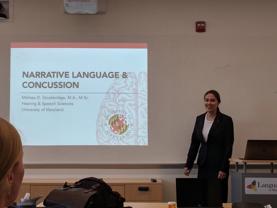 We’re all very proud of Melissa Stockbridge, who successfully defended her dissertation, “The role of personality on cognitive-linguistic deficits in teens and adults with mild TBI.” Dispositional negativity — here indexed using BFI-2-N — strongly predicts patient reports of symptoms in the months following a concussion. In fact, Melissa showed that this is true even if you censor overlapping content. In contrast, patient disposition was unrelated to computerized measures of cognitive function. Collectively, these results suggest that everyday clinical decision-making (e.g. return to play) can be biased by patient personality, and underscore the promise of developing clinic-friendly e-cog technology for objective testing. A key challenge for the future will be to prospectively assess these personality-symptom relations in high-risk individuals (e.g. student-athletes, combat personnel). Congratulations, Dr. Stockbridge!
Melissa Stockbridge will be defending her dissertation. Her presentation is open to all.
The role of personality on cognitive-linguistic deficits in teens and adults with mild TBI
Thursday, May 17, 2018 – 2:00pm to 4:00pm
Language Science Center; room 2124 HJ Patterson Hall
Even the mildest form of traumatic brain injury, concussion, can result in adverse physical, cognitive, behavioral, and social consequences. Concussion injuries frequently result in patients who describe deficits in daily communication and overall “fogginess,” but whose deficits are not consistently captured on traditional assessments of language. The purpose of this research was two-fold: first, to examine typed written communication in order to better understand the kinds of cognitive and language deficits that adolescents and adults experience immediately and chronically following a concussion; and second, to examine the influence of a particular trait-like dimension of personality and temperament, the propensity toward more frequent, intense, and enduring negative affect (called dispositional negativity), on exacerbation of these deficits. Using a survey conducted entirely online, 92 participants aged 12-40 years old who had a recent concussion, a history of concussion, or no history of brain injury wrote two narrative samples and an expository sample, completed multiple tasks targeting word-level and domain general cognitive skills, and provided rich self-report information important to better understanding their personality, temperament, and mental health. Performance by recently injured participants suggested that deficits in narrative language, though likely influenced by problems in word-finding, memory, and attention, also existed beyond what could be explained by those deficits alone. Narrative-specific deficits were observed in written content, organization, and cohesiveness. Moreover, including dispositional negativity in models of concussion history (group) and self-reported somatic symptomology improved the sensitivity and specificity of these models, which supports the value of considering individual differences in personality when engaged in concussion management.


Gloria Kim has been selected for a Maryland Summer Scholars Award from the Maryland Center for Undergraduate Research. Congratulations! Her project, which is mentored by Juyoen Hur, a postdoctoral fellow in the lab, builds on our on-going NIMH sponsored study focused on understanding the brain systems that confer risk for developing mood and anxiety disorders in university students.
Dr. Shackman has joined the Editorial Board of Neuropsychologia as Emotion and Social Neuroscience Section (Handling) Editor.
As part of our lab’s on-going commitment to promoting reproducible science, accelerating the pace of scientific discovery, and maximizing the impact of public support for biomedical research, we are sharing data from several recent imaging studies via NeuroVault.org. Links to the relevant data collections can be found in the Publications section of our website.
Please join us for student presentations at SFN/DC and ABCT/San Diego!
SFN
Melissa Stockbridge (UMD/Newman Lab) will be presenting recent work using a combination of brain imaging techniques—including high-precision single-subject analyses and machine learning-based brain signatures—to demonstrate that anxiety, pain, and cognitive control are functionally integrated in the mid-cingulate cortex (MCC) at SFN on Wednesday November 15, 2017, 8:00 – 12:00 PM Halls A-C as part of Session 700 – Emotional States: Empathy. 700.09 / PP28. This work is part of a collaboration with Luiz Pessoa, Dave Seminowicz, Tor Wager, and Wani Woo.
ABCT
Allegra Anderson and Katie DeYoung will be presenting new experience-sampling (EMA) work demonstrating that individuals who, by virtue of their more negative/neurotic disposition, are at risk for developing anxiety disorders and depression are actually more responsive to the ‘mood brightening’ impact of positive daily events. Matt Barstead (UMD/Rubin Lab) was also involved in the project. Allegra will be presenting the poster at ABCT on (PS11- #A31) on Saturday, November 18 12:15 PM – 1:15 PM Location: Indigo Ballroom CDGH, Level 2, Indigo Level.
Jim Doorley (GMU/Kashdan Lab) will be describing work aimed at better understanding the momentary experience of social anxiety disorder at ABCT on Saturday, November 18 2:00 PM – 3:30 PM Location: Aqua Salon A & B, Level 3, Aqua Level.
If you’re interested in learning more about our work, please join us for upcoming presentations–
Department of Psychological and Brain Sciences, University of Iowa: October 27, 2017
S4SN Meeting, DC: November 10, 2017. Join us for a wonderful symposium organized by Kalina Michalska!
SFN Meeting, DC: November 15, 2017. Melissa Stockbridge will be presenting some exciting new collaborative work focused on the integration of anxiety, pain, and cognitive control in the cingulate!
Department of Psychology Neuroscience Seminar Series, Yale University: November 17, 2017
Department of Psychology, Vanderbilt University: March 29, 2018
ADAA Meeting, DC: April 6-7, 2017. Join us for a symposium on recent advances in our understanding of extreme early-life anxiety, featuring Jenni Blackford (Vanderbilt), Dylan Gee (Yale), and Lisa Williams (Wisconsin)!
“Neural circuitry governing anxious individuals’ mis-allocation of working memory to threat,” an article co-authored by Dr. Alex Shackman, was featured on Psychpost!
The article details how threatening information invades the working memory of anxious individuals. Check out the story here!
Allegra has been chosen to attend the University of Minnesota’s Diversity in Psychology Program! This is a competitive award granted to historically underrepresented individuals in psychology graduate programs who are interested in learning doctoral training at UMN.
The program involves meetings with program faculty members and graduate students to discuss students’ research interests and presentations designed to familiarize participants with strategies for constructing successful graduate school applications. Travel, lodging, and meals are provided for all attendees.
Congrats, Allegra!
The Shackman lab is thrilled to have joined the Hierarchical Taxonomy of Psychopathology (HiTOP) Consortium, an international workgroup of clinical and personality psychologists, psychiatrists, statisticians, and neurobiologists focused on understanding the nature of mental illness and using data-driven approaches to develop better and more useful tools for diagnosing and measuring psychiatic disorders and symptoms.
You can learn more about HiTOP here.
Samiha recently presented her undergraduate honors thesis project in a poster session at the 125th Annual Convention of the American Psychological Association (APA) in Washington, D.C. Her thesis explored risk factors for psychological well-being and distress among South Asian American young adults.
APA is the largest annual gathering of psychologists from across the world, featuring individuals from a variety of professional settings presenting on topics from all sub-fields in psychology. This organization’s main purpose is to communicate methods of improving health and wellness through the practice of psychology.
Congrats, Samiha!
Allegra has been invited to the University of Delaware’s Clinical Psychology Doctoral Program Visit Day! This is a competitive award granted to research-oriented students and recent graduates from underrepresented groups who want to learn more about doctoral training in clinical science.
The program involves informative presentations on applying to graduate school, individual and small group meetings with program faculty members and graduate students to discuss students’ research interests, and social events & networking with current graduate students and faculty members. Travel, lodging, and meals are provided for all attendees.
Congratulations, Allegra!
A very warm welcome to our new post-doctoral fellow, Juyoen Hur!
Juyoen is a postdoctoral fellow in the Department of Psychology at the University of Maryland, College Park. She earned her Ph.D. in Clinical Psychology from the University of Illinois at Urbana-Champaign in 2017 and completed her clinical internship at the Department of Psychiatry of the University of Wisconsin-Madison School of Medicine and Public Health. Her program of research examines the attentional and affective mechanisms of anxiety disorders and depression, particularly focusing on the interactive influences of emotion and attention. Her interest in studying the interplay of cognitive and emotional risk factors for anxiety and depression stems from its potential to identify novel treatment targets and thus improve prevention and intervention efforts.

Claire is currently completing a clinical externship with the Health Psychology and Neurobehavioral Research Group at the National Cancer Institute within NIH. Her training focuses on neuropsychological assessment and treatment of the cognitive, behavioral, and psychological problems of adolescents and adults with chronic medical conditions, especially those with neurobehavioral effects (e.g., brain tumors, leukemia, sickle cell anemia, neurofibromatosis type 1), enrolled on treatment protocols at the NCI and other NIH institutes.
Rachael’s current clinical externship placement is at the Center for Autism Spectrum Disorders at Children’s National Hospital. Her training focuses on neuropsychological and diagnostic evaluations for children, adolescents, and young adults on the autism spectrum, or suspected of having an autism spectrum disorder. Go, Rachael!
Claire successfully defended her master’s project.
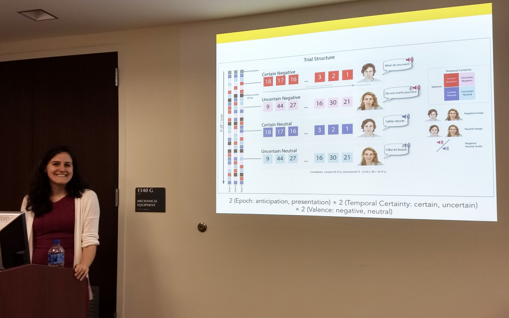
This functional MRI project is focused on understanding the anxiety-reducing effects of alcohol on the neural systems underlying fear and anxiety in a community sample of healthy adults (21-35 years). For this project, we developed and employed a novel fMRI task paradigm which selectively manipulates temporal certainty during threat-of-shock.
In particular, Claire’s masters focused on the dose-dependent consequences of acute alcohol intoxication on the two major divisions of the central extended amygdala–the bed nucleus of the stria terminalis and the central nucleus of the amygdala.
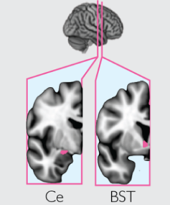
This work builds on prior mechanistic studies in rodents and psychophysiological findings in humans, and helps set the stage for more integrative and bidirectional translational models to elucidate mechanisms underlying anxiety and substance abuse.
The supervising committee consisted of Drs. Alex Shackman, Luiz Pessoa, Ed Bernat, and Matthew Roesch.
Congrats, Claire!
The Shackman lab is out and about! Please join us for what promises to be a fun and interesting series of talks and symposia!
The 2016-17 UMD combined colloquia series has been amazing, providing members of the lab with a number of outstanding opportunities for discussing our work with outside experts, including Jonathan Fadok, Katia Harle, Sabine Kastner, Hedy Kober, Liz Phelps, Russ Poldrack, Kerry Ressler, and Lucina Uddin. Still to come, presentations from Todd Braver and Michael Platt.
In addition, a number of outstanding scientists will be descending on Baltimore next week for the annual Maryland Neuroimaging Retreat, including Lauren Atlas, Catherine Bushnell, Cam Craddock, Martin Lindquist, Ben Seymour, Tor Wager and our very own, Alex Shackman. Please consider joining us for what promises to be an exciting series of talks and panel discussions.
And next year: Kay Tye!
Candidates are being considered for a NIMH-funded postdoctoral position in the laboratory of Dr. Alex Shackman in the Department of Psychology at the University of Maryland at College Park (http://shackmanlab.org/). The overarching mission of the lab is to have a deep impact on the fields of affective and translational neuroscience. To that end, we do our best to perform innovative studies that can lead to significant discoveries, to disseminate our discoveries as widely as possible, and to mentor trainees to become top-notch scientists. As part of a recently awarded R01 (MH107444), the major focus of this position will be on understanding the neural circuitry underlying fear and anxiety and its role in the development of anxiety disorders, depression, and substance abuse in young adults. A secondary focus will be on linking variation in the function of that circuitry to thoughts, feelings, and behavior in the real-world, indexed using ecological momentary assessment (EMA) techniques. Eye-tracking measures of attention and peripheral physiological measures of arousal will also be incorporated. There will be opportunities to become involved in other projects and to develop new analytic strategies. We are particularly excited about candidates with a strong background in fMRI methods, but will also consider those with expertise in other areas of affective neuroscience or clinical psychology. Applicants should have a Ph.D. in a relevant field; strong publication record; expertise in human cognitive, affective, or clinical/translational neuroscience; and excellent organizational and interpersonal skills. This is an excellent opportunity for receiving top-notch mentorship in affective/translational neuroscience in a highly productive environment. This is a 1 year position that is renewable for a total of 3 years, contingent on performance and funding. Applicants should send a cover letter describing relevant experience and interests, CV, and contact information for 3 references to Dr. Shackman (shackman@umd.edu). Applicants will be considered until the position is filled. The University of Maryland is an Equal Opportunity/Affirmative-Action Employer.
Read more about the project here and here.
Study Coordinator
Post-baccalaureate Position in Affective and Clinical/Translational Neuroscience
University of Maryland, College Park, MD
Candidates are being considered for a NIH-funded post-baccalaureate (study coordinator) position in the laboratory of Dr. Alex Shackman in the Department of Psychology at the University of Maryland at College Park (http://shackmanlab.org/). The overarching mission of the lab is to have a deep impact on the fields of affective and clinical/translational neuroscience. To that end, we do our best to perform innovative studies that can lead to significant discoveries, to disseminate our discoveries as widely as possible, and to mentor trainees to become top-notch scientists. As part of several recent NIH awards, the focus of this position will be to support on-going projects aimed at understanding the neurobiology of fear and anxiety and its role in the development and maintenance of adult anxiety disorders, depression, and substance abuse. This position will provide opportunities to gain experience with neuroimaging (fMRI), ecological momentary assessment (EMA), eye-tracking, psychophysiological, and clinical interviewing techniques. This is an exciting opportunity for receiving top-notch mentorship and establishing a competitive research record in preparation for graduate school. This is a 1 year position that is renewable for a total of 2 years, contingent on performance and funding. Duties may include, but are not limited to, subject recruitment, data acquisition using behavioral and neuroimaging techniques, study management, database management, data processing/analysis, and lab management. We are particularly excited about candidates with a background in imaging methods, programming, digital signal processing, quantitative methods, statistics, or related areas. Applicants should send a cover letter describing relevant experience and interests, CV/resume, and 2-3 letters of reference to Dr. Shackman (shackman@umd.edu). Applicants will be considered until the position is filled. The University of Maryland is an Equal Opportunity/Affirmative-Action Employer.
Read more about the NIMH R01 project here and here. Read more about the NIDA R21 project here and here.
Congratulations to Rachael Tillman, who passed her qualifying exams to become an official doctoral candidate in clinical psychology!
Richard Hum, an RA in the lab, was recently accepted into the Biology Honors Program, joining a long list of lab RA’s who have participated in similar campus programs, including the Biological Sciences Honors Internship Program, Integrated Life Sciences (ILS) Honors Program, Research Internship in Science and Engineering (RISE) Scholarship Program, and Summer Research Initiative (SRI) Fellowship Program. Congrats, Richard!

Congratulations to graduate student Rachael Tillman, who received a fellowship that will enable her to attend the upcoming Tools of the Trade Workshop at UCLA. The workshop is jointly sponsored by the National Institutes of Health and Stanford Center for Reproducible Neuroscience. Read more about the imaging workshop here.
Rachael successfully defended her master’s project.
This functional MRI project is focused on clarifying the architecture of functional architecture of circuits centered on the two major divisions of the central extended amygdala–the bed nucleus of the stria terminalis and the central nucleus of the amygdala–in a large sample of healthy, community dwelling adults. This work lays a basic neuroscience foundation for understanding alterations of these circuits in psychiatric populations.
The supervising committee consisted of Drs. Alex Shackman, Elizabeth Redcay, Luiz Pessoa, and Nathan Fox.
Congrats, Rachael!
Slides and syllabi are now available for:
Updated for Fall 2016.
We are now accepting applications for the 2017 in-coming class. You are welcome to apply via the Clinical area group in the Department of Psychology or via the Neuroscience and Cognitive Science (NACS) training program. Learn more here: http://shackmanlab.org/positions/graduate-students/
Please note that the major focus of this position will be on neural circuitry underlying fear and anxiety in young adults. A secondary focus will be on linking variation in the function of that circuitry to thoughts, feelings, and behavior in the real-world, indexed using ecological momentary assessment (EMA) techniques. Eye-tracking measures of attention and peripheral physiological measures of arousal will also be incorporated. We are particularly excited about candidates with a passion for fear/anxiety and a strong background in imaging, programming, digital signal processing, or related areas.
This research is funded by the University of Maryland and the National Institutes of Health.
Thanks to generous support from the National Institutes of Health, lab members now have access to a total of 4 servers (Badger, Ferret, Fisher, and Polecat), which will enable us to keep abreast with a growing flood of new imaging and peripheral physiological data.

If you’re curious, you can read more of the details here.
Congratulations to Claire Kaplan, who was honored with a travel award by the Society for Research in Psychopathology for her poster entitled ‘Understanding the neurobiology of fear and anxiety.’ This a huge honor only given to the top 5% of posters at the meeting. Claire’s project leverages a combination of multiband functional MRI and a novel MultiThreat Countdown task to characterize the neural systems engaged by certain and uncertain threat.
You can view the poster here and read some related papers here and here. The task was adapted from earlier fear-potentiated startle work by Hefner, Curtin and colleagues.
This work was supported by the University of Maryland, NSF, and NIH.
Dr. Shackman will be speaking at:
56th Annual Meeting of the Society for Psychophysiological Research (SPR): September 21-25 (Time/Room TBA), 2016, at the Marriott City Center Hotel, Minneapolis
UMD Counseling Center: November 30, 2016 at noon.
The lab provides an excellent opportunity to get hands-on experience with data collection. It’s a great way to prepare for successful careers in research (graduate school, postbac fellowships at the NIH and Lieber Institute, NSF REU fellowships) or medicine (medical school)!
Learn more about the lab’s philosophy and mission here. Or, explore our website to find out more about the exciting kinds of research conducted by our team.
In order to apply, please download the application form. Email the completed application and an unofficial copy of your transcripts to Dr. Shackman (shackman@umd.edu).
Congrats to Claire!
Claire Kaplan’s poster focused on “Understanding the neurobiology of fear and anxiety” was selected as one of the preliminary finalists for the Smadar Levin Student Poster Award by the Society for Research in Psychopathology (SRP). Congratulations!
To learn more about SRP, please visit their website.
Shackman lab receives NIDA R21 award
The National Institute of Drug Abuse (NIDA) has awarded a $418,000 grant to the University of Maryland to support research aimed at understanding the neurobiology of nicotine dependence.
Nearly 50 million Americans smoke tobacco and smoking is the leading cause of premature death and disability in the US. The grant will support a team of researchers, led by Professor Alex Shackman in the Department of Psychology at Maryland, who plan to use state-of-the-art brain imaging techniques and smart-phone technology to clarify, for the first time, the relevance of anxiety-related brain circuits to chronic tobacco use and acute nicotine withdrawal in humans—a critical step toward the development of new, brain-based cessation aids.
Dr. Shackman notes that, “while the transition from tobacco use to nicotine dependence is associated with long-lasting changes in multiple emotional and motivational mechanisms, most neurobiological research in humans has focused on reward-related systems. This is unfortunate because heightened anxiety is a hallmark of nicotine deprivation and there is compelling evidence that anxiety and negative affect powerfully motivate nicotine dependence and relapse.”
The investigators plan to use functional MRI (fMRI) to quantify anxiety-related brain function in abstinent and non-abstinent tobacco smokers. Mobile phone technology will be used to assess smoker’s daily perceptions of stress, anxiety, depression, and craving, as well as actual smoking behavior. According to Dr. Shackman, this project “will provide an important first opportunity to see whether models of addiction developed in rodents apply to humans and hopefully will inform the development of new treatments for tobacco addiction.”
Other members of the investigative team include Dr. Jason Smith in the Department of Psychology at Maryland; Dr. Luiz Pessoa, Director of the Maryland Neuroimaging Center; Dr. Megan Piper from the Center for Tobacco Research and Intervention at the University of Wisconsin-Madison; and Dr. John Curtin in the Department of Psychology at the University of Wisconsin-Madison.
7/11/2016 | College Park, MD USA
The National Science Foundation has awarded a prestigious Graduate Research Fellowship to Claire Kaplan, a second year graduate student in the laboratory. This will provide three years of support for her exciting program of neuropsychopharmacology research.
It’s common knowledge that a drink or two reduces anxiety. While the molecular underpinnings of this anti-anxiety effect is well established, remarkably little is known about the brain networks that mediate the anxiety-reducing effects of alcohol or other widely used anxiolytic medications. Claire’s work aims to address this fundamental question. Her on-going research is focused on establishing the influence of different doses of alcohol on the function of brain circuits implicated in fear and anxiety. Another project, still in design stages, will focus on the impact of the benzodiazepines, a drug commonly prescribed for patients suffering from extreme anxiety. This program of research promises important new insights into the mechanisms that support pathological anxiety and contribute to the development of addiction. Ultimately, it promises to inform the development of more precise treatments for a range of common, often debilitating mental illnesses.
Claire’s research builds on the strengths of an international team of scientific collaborators, including Dr. Jason Smith, in the Department of Psychology at Maryland; Dr. Luiz Pessoa, Director of the Maryland Neuroimaging Center; Dr. John Curtin, in the Department of Psychology at the University of Wisconsin; and Dr. Siri Leknes, Oslo University Hospital, Oslo, Norway.
3/30/2016 | College Park, MD USA
The National Institute of Mental Health (NIMH) has awarded a 3.4 million dollar grant to the University of Maryland to support research aimed at understanding the mechanism that that promote the development of pathological anxiety and depression.
A growing body of data shows that these disorders impose a staggering burden on public health and the global economy, making them a growing concern for clinicians, researchers, and public policy makers. The anxiety disorders are the most common family of mental illnesses in the US and Europe and often contribute to the development of depression and substance abuse. Existing treatments are inconsistently effective or associated with significant side effects.
The new grant will support an international team of researchers, led by Professor Alex Shackman in the Department of Psychology at Maryland, who plan to use state-of-the-art brain imaging techniques, clinical measures, and smart-phone technology to clarify the mechanisms that support to the development and recurrence of anxiety disorders and depression—a critical step toward the development of new, brain-based strategies for preventing or treating these illnesses.
“This is a really exciting project, notes Dr. Shackman, “These disorders contribute to the suffering and misery of millions of patients and their loved ones all over the world, including many students at Maryland and other universities. The pipeline for developing new drugs is stalled. It’s imperative that we identify the brain circuits that underlie extreme anxiety and get a better handle on their relevance to changes in mood and function in the real world, close to the kinds of end-points that patients, families, and clinicians care about.”
One of the goals of the project is to use smart-phone technology to sample feelings, behavior, stress, and social support in college freshmen. “This is really one of the most novel and exciting parts of the project,” says Dr. Shackman. “I think the research community has done a great job using brain imaging techniques in people and more mechanistic interventions in rodents and monkeys to identify candidate circuits, networks in the brain that control anxiety. But we know almost nothing about whether any of those circuits actually predict anxiety, depression, or stress-reactivity where it matters, out in the real world. Here, we plan to use the smart-phones to intensively sample our subjects’ daily experience repeatedly for 30 months, as they transition from freshman year, to sophomore year, to junior and even senior year of university. We can then link those measures of experience and behavior back to measures of brain function collected during their freshman year, which we hope will enable us to discover patterns of brain function that predict who gets sick, who gets depressed, who starts drinking by themselves to cope with their stress.”
These data may provide new targets for drug development and guide the development of improved animal models of mental illness. “And it’s not just drugs,” Shackman emphasized, “this might give us some really important new clues about ways to better identify high-risk individuals before they get sick, and guide them into prevention programs focused on developing more effective coping skills. And the smart phone data may help us to develop better mobile apps for treating or even preventing these disorders, before relationships are strained, before performance in school or the workplace really starts to suffer.”
Other members of the international investigative team include Dr. Jason Smith in the Department of Psychology at Maryland; Drs. Greg Hancock and Nathan Fox in the Department of Human Development and Quantitative Methodology at Maryland; Dr. Luiz Pessoa, Director of the Maryland Neuroimaging Center; Dr. Todd Kashdan in the Department of Psychology at George Mason University; and Dr. Matthias Gamer in the Department of Psychology at the University of Würzburg, Germany.
To learn more about anxiety disorders and depression, please visit the Anxiety and Depression Association of America website.
3/30/2016 | College Park, MD USA
Recent research from Dr. Shackman and colleagues at the Center for Investigating Healthy Minds has been incorporated into the Spring 2016 ‘Fuel Happiness’ campaign organized by Lululemon, the international athletic apparel company.

In a project spearheaded by Dr. Helen Weng, we demonstrated that compassion training can increase prosocial behavior and is associated with changes in a complex network of brain regions involved in social cognition and emotion regulation. This work provides preliminary scientific evidence that compassion can be systematically trained and that increased altruism emerges from changes in the brain.
The Fuel Happiness campaign is designed to spread the news about recent meditation and mindfulness research and, in so doing, encourage all of us to to take a few moments to cultivate an increased sense of compassion for themselves and others.
Weng, H. Y., Fox, A. S., Shackman, A. J., Stodola, D. E., Caldwell, J. Z. K., Olson, M. C., Rogers, G. M. & Davidson, R. J. (2013). Compassion training alters altruism and the neural responses to suffering. Psychological Science, 24, 1171-80.
Learn more about the paper here.
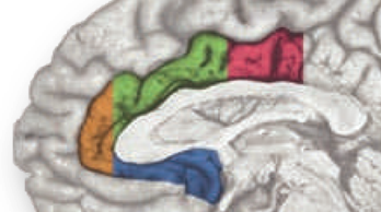
The rostral cingulate cortex—the thick belt of tissue encircling the corpus callosum—plays a central role in contemporary models of emotion, pain, and cognitive control (https://tinyurl.com/hvld5yd). Work in these three basic domains has, in turn, profoundly influenced contemporary perspectives on more complex phenomena, including social cognition and a variety of neuropsychiatric disorders (https://tinyurl.com/neb48x9).
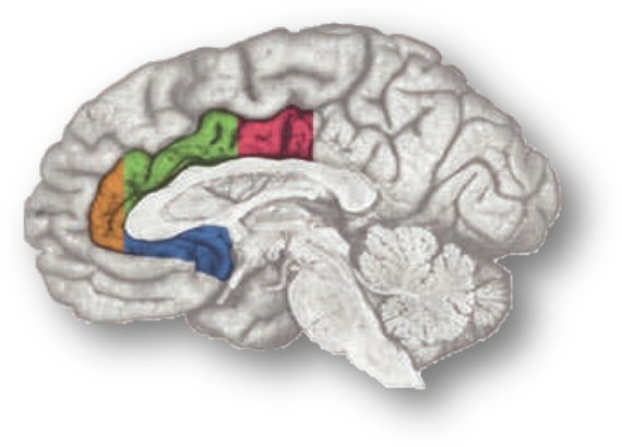
Despite this progress, the functional architecture and significance of activity in the rostral cingulate cortex remains enigmatic. We still don’t have a definitive answer to the question, What exactly does the cingulate ‘do’?
In a recent high-profile report (https://tinyurl.com/ofnxhtp), Matt Lieberman and Naomi Eisenberger make 3 extraordinary claims about the function of the dorsal or mid-cingulate cortex (‘dACC/MCC’), the region shown in green and magenta region in the accompanying figure:
1. We’re labeling it incorrectly. Lieberman and Eisenberger suggest that a substantial number of prior reports mis-labeled activation clusters lying in the more dorsal supplementary motor area (SMA) and pre-SMA as dACC/MCC:
“studies focused on the dACC are more likely to be reporting SMA/pre-SMA activations than dACC activations.”
2. We’re asleep at the wheel—Cognitive control does not reflect dACC/MCC. Lieberman and Eisenberger claim that standard lab assays of cognitive conflict and control—including the widely used Stroop, Eriksen flanker, Go/No-Go, and Stop signal tasks—do not produce robust activation in the dACC/MCC, contrary to more than a decade’s worth of imaging studies…
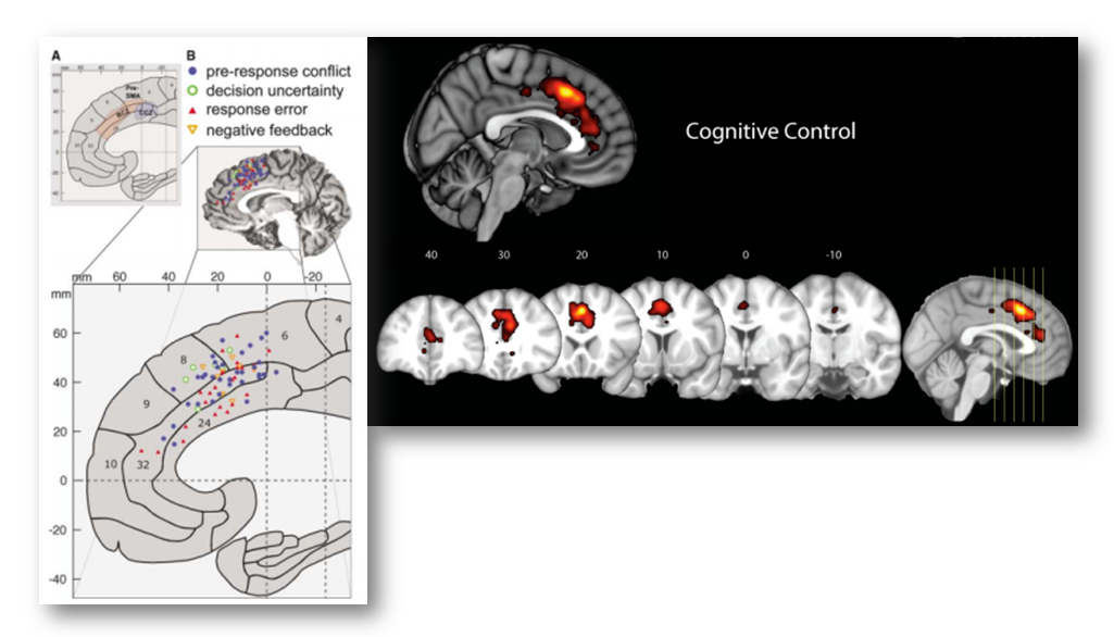
As they note,
“for many of the terms, the lion’s share of the activity is…in the [more dorsal] SMA or pre-SMA [i.e. the region depicted in green]…[Indeed] the forward inference maps for…“stop signal,” [and] “Stroop” each have a relatively modest footprint [in the dACC/MCC proper; the region depicted in blue].

3. The dACC/MCC is selective for pain. Based on automated meta-analyses of hundreds of brain imaging studies—performed using tools available at Neurosynth.org—Lieberman and Eisenberger argue that:
“The only psychological phenomenon that can be reliably inferred given the presence of dACC activity is pain…The conclusion from the Neurosynth reverse inference maps is unequivocal: The dACC is involved in pain processing. When only forward inference data were available, it was reasonable to make the claim that perhaps dACC was not involved in pain per se, but that pain processing could be reduced to the dACC’s “real” function, such as executive processes, conflict detection, or salience responses to painful stimuli. The reverse inference maps do not support any of these accounts that attempt to reduce pain to more generic cognitive processes. As seen in Fig. 3, a whole slew of such terms show little or no evidence of being likely candidates to explain the psychological bases of dACC activity. Although some of these functions might be instantiated in the [more dorsal] SMA or pre-SMA, they do not seem to be reliably related to dACC activity”
Naturally, such extraordinary, high-profile claims are bound to attract critics and skepticism. One of the most damning critiques comes in the form of a series of blog posts written by Tal Yarkoni, the developer of the software that Lieberman and Eisenberger used for their analyses (https://tinyurl.com/nvt3vlr and https://tinyurl.com/hhmgxvu). The posts are thoughtful, comprehensive, at times cranky, and well worth reading in their entirety, but for Yarkoni the bottom line is that:
“No, the dorsal anterior cingulate is not selective for pain….Lieberman & Eisenberger (2015) argue, largely on the basis of evidence from my Neurosynth framework, that the dACC is selective for pain. They are wrong. Neurosynth does not—and, at present, cannot—support such a conclusion. Moreover, a more careful examination of Neurosynth results directly refutes Lieberman and Eisenberger’s claims, providing clear evidence that the dACC is associated with many other operations, and converging with extensive prior animal and human work to suggest a far more complex view of dACC function.”
In particular, Yarkoni notes that voxels in the dACC/MCC are likely to be recruited in studies of fear, cognitive control, and conflict (see the following figure adapted taken from his post). If you’re curious, you can generate and download the same reverse inference maps yourself over at www.Neurosynth.org.

Given these observations, Yarkoni concludes that,
“In every single one of these cases, we see significant associations with dACC activation in the reverse inference meta-analysis. The precise location of activation varies from case to case…but the point is that pain is clearly not the only process that activates dACC. So the notion that dACC is selective to pain doesn’t survive scrutiny even if you use [Lieberman and Eisenberger’s] own criteria.”
For what it’s worth, I agree with Yarkoni’s conclusion, which he arrives at entirely based on a consideration of the statistics (see especially his second post, https://tinyurl.com/hhmgxvu).
A neuroanatomical perspective on the dACC/MCC
My own concern with Lieberman and Eisenberger’s study—one not addressed in either Yarkoni’s critique or Lieberman and Eisenberger’s thoughtful rebuttal (https://tinyurl.com/ptvhmjm)—has nothing to do with meta-analysis, Bayes, or statistics per se.
From my perspective, the crux of this story comes down to neuroanatomy—What exactly is the dACC/MCC?—Which voxels should be considered ‘in’ and which should be left ‘out’?
The answer to this fundamental question is central to Lieberman and Eisenberger’s claim that cognitive conflict and control engage the SMA/pre-SMA, whereas pain (and perhaps related states of fear and anxiety) engage the dACC/MCC. To foreshadow my conclusion, in the remainder of this post I will describe why Lieberman and Eisenberger’s approach to identifying the dACC/MCC is problematic and how this undermines their remarkable inferences.
My motivation—It’s about the cingulate, not Lieberman & Eisenberger
As an aside, I want to briefly address my motives for joining the public conversation about Lieberman and Eisenberger’s paper.
First, in the spirit of transparency, my colleagues and I have invested considerable effort trying to understand the rostral cingulate. If you’re curious, you can read more about our take here (https://tinyurl.com/hpqmkwn) and here (https://tinyurl.com/np3d3nb). In brief, we have hypothesized that the dACC/MCC represents a hub, where information about punishment, errors, conflict, and other kinds of negative feedback is integrated and used to bias or control our thoughts, feelings, and actions in the face of uncertainty about actions and motivationally significant outcomes, such as punishment. One of the motivations for this hypothesis was evidence from imaging studies and neuronal recordings that tasks that elicit negative affect (e.g. fear), physical pain, or the need for increased cognitive control all recruit an overlapping territory in the anterior dACC/MCC.
Naturally, Lieberman and Eisenberger’s claim that dACC/MCC activation is specific to pain seems to run counter to the idea that this key brain region is consistently recruited across diverse psychological states. But, in their rebuttal to Yarkoni, Lieberman and Eisenberger clarify their conclusions in ways that ameliorate some of my initial concerns, arguing for example that they simply meant that “pain is a more reliable source of dACC activation than the other [i.e. cognitive] terms of interest” and that the dACC/MCC is, in fact, exquisitely sensitive to negative affect: “We have long posited that one of the functions of the dACC was to sound an alarm when certain kinds of conflict arise. We think the dACC is evoked by a variety of distress-related processes including pain, fear, and anxiety.” Still, their claims that the dACC/MCC is only weakly sensitive to demands for increased cognitive control was at odds with my own understanding of the literature and my interpretation of the maps that I could generate with a few button clicks in Neurosynth. So, I wanted to understand why we were reaching such different conclusions.
My second motive for entering this debate is more substantial and, hopefully, generative. I want to understand what the dACC/MCC does and leverage that understanding to guide the development of more effective interventions. My concern is that there is widespread misunderstanding about what constitutes the dACC/MCC. For example, in their blog posts, Lieberman, Eisenberger, and Yarkoni all seem to agree that the cluster in this next figure does not lie in the dACC/MCC—but based on the evidence that I review below, I believe that this is incorrect.

I hope that by reviewing what is known about the anatomy of the dACC/MCC and articulating some simple, concrete methodological recommendations for future imaging studies, it will accelerate our understanding; that the graduate students, post-docs, and PI’s who attempt to build on the published literature will adopt methods that permit stronger inferences about what the cingulate does.
Lieberman and Eisenberger fail to address individual variation in anatomy
Lieberman and Eisenberg describe the dACC/MCC as the
“section of the cingulate that sits above the corpus callosum…the cingulate sulcus is the dorsal boundary of the dACC. Above this sulcus are the supplementary and pre-supplementary motor areas (SMA and pre-SMA), which…are contained within the superior frontal gyrus.”
This is the only description in the paper of how they prescribed their dACC/MCC region-of-interest (ROI; the blue region in the next figure). Fortunately, we can reverse engineer what they probably did. Close inspection of their figures suggests that the dACC/MCC ROI was manually prescribed on the Colin27 brain (https://tinyurl.com/o55nffe; left panel below), which is simply the average of 27 anatomical scans of a single adult man (the eponymous Dr. Colin J. Holmes, shown in the right panel) in standard stereotaxic space (https://tinyurl.com/q2jqgeu).

What’s wrong with relying on the brain of one Canadian scientist for drawing the dACC/MCC ROI? After all, they’ve given us Hebb, Penfield, the MNI, and Tim Horton’s right? Well, as the developers of the Colin27 template note:
“this dataset was not originally intended for use as a stereotaxic template… As a single brain atlas, it did not capture anatomical variability and was, to some degree, a reversion to the [outdated] Talairach approach.”
In other words, Lieberman and Eisenberger conducted meta-analyses based on hundreds of brain imaging studies, collectively encompassing thousands of individual subjects, each with their own idiosyncratic brain.Each one of those thousands of brains was ‘warped’ (i.e. ‘registered’ or ‘normalized’) to a standard brain template by the original investigative teams. This standardization not only allowed those investigators to compute group statistics, it‘s also what makes it possible to conduct large-scale meta-analyses in Neurosynth or other software packages (http://brainmap.org)—all of the activation peaks are reported in the same 3D Cartesian space (more or less; https://tinyurl.com/hjqy6r5).
The problem is that the warping process is imperfect; there are errors in how well each brain is registered to the template, especially in studies relying on earlier generations of warping techniques (like the well-known Talairach and Tournoux stereotaxic technique; https://tinyurl.com/o83ksuk). So, it’s not appropriate to think of features of the brain—like the dACC/MCC—as a crisp point or line, as in the left panel of the following figure.

Given registration errors across many, many individual brains, it’s better to think of these features in a more probabilistic way (as in the right panel above), which is why most investigators (and NeuroSynth) use brain templates that were generated by averaging across tens or hundreds of warped brains, as in the popular MNI152 template shown below (https://tinyurl.com/zgjv4a3).

In sum, the Colin27 brain template (apparently) used by Lieberman and Eisenberger conveys a false sense of precision about the sulcus separating the cingulate from the overlying SMA/pre-SMA. But this is only the first part of what is really a compound problem. As it turns out, we probably do not want to rely on just the cingulate sulcus, let alone just Colin’s cingulate sulcus.
Lieberman and Eisenberger define the dACC/MCC in an arbitrary manner
It’s worth pausing for a moment and considering how the rostral cingulate has traditionally been defined on the basis of invasive studies of cellular morphology and chemistry. The neuroanatomist Korbinian Brodmann (https://tinyurl.com/glex9gp) first defined the rostral cingulate as the complex formed by architectonic areas 24, 32, 25, and 33. Of these, areas 24, 32, and 33 (marked with red circles below) constitute the subdivision lying dorsal to the corpus callosum (dACC/MCC).
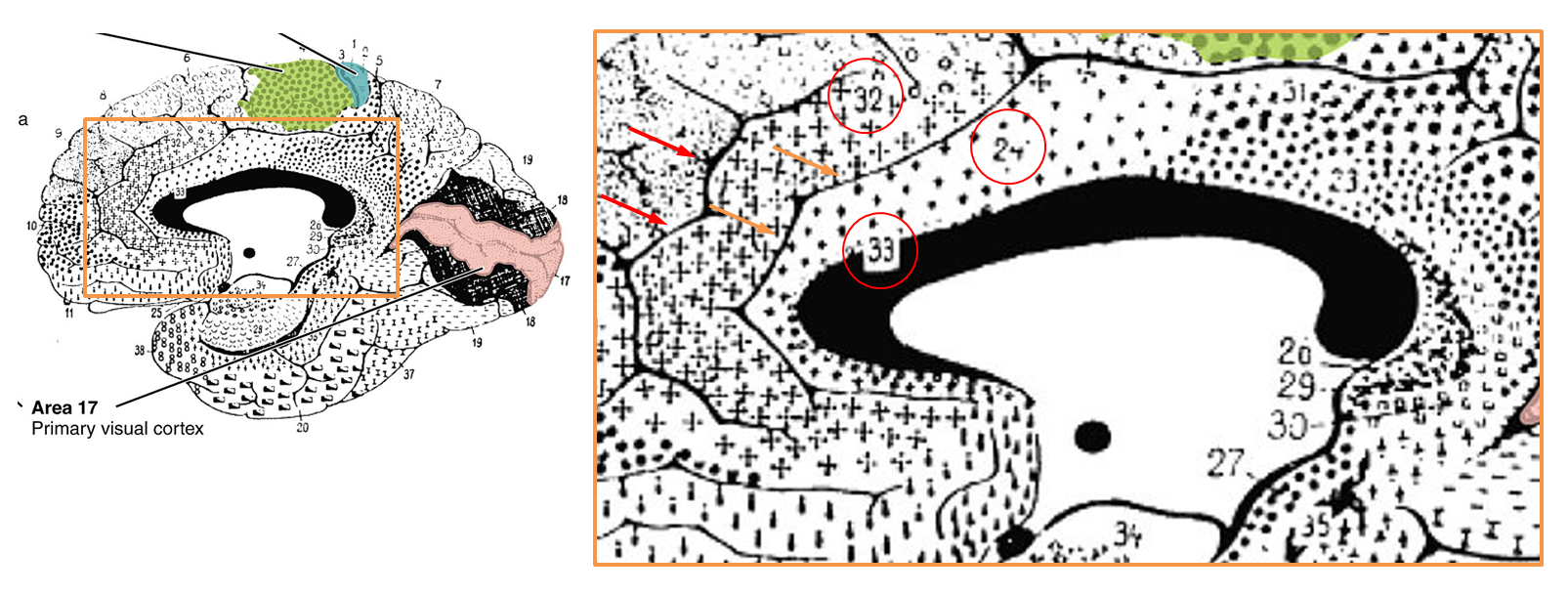
As shown in the next figure, much of the cortical gray matter that makes up the dACC/MCC lies buried in sulci and is not visible in sagittal images. For brains—like Colin’s or that shown in the next figure—that only have a single cingulate sulcus, Lieberman and Eisenberger’s dACC/MCC ROI probably does a pretty good job. As shown in upper right panel (https://tinyurl.com/hvld5yd), as long as they included the voxels located in the dorsal bank of the cingulate sulcus (shown in purple), their ROI would encompass all of the dACC/MCC (i.e. areas 33, 24, and 32).

Whether that’s the case is unknown; the ROI protocol is not described in Lieberman and Eisenberger’s report and they do not show any coronal images. It’s not really any more clear in their rebuttal to Yarkoni, although it looks like they may be missing some of the dorsal bank (technical aside: the green line demarcating the dACC/MCC region is not a genuine 3D ROI and is clearly for illustrative purposes only).
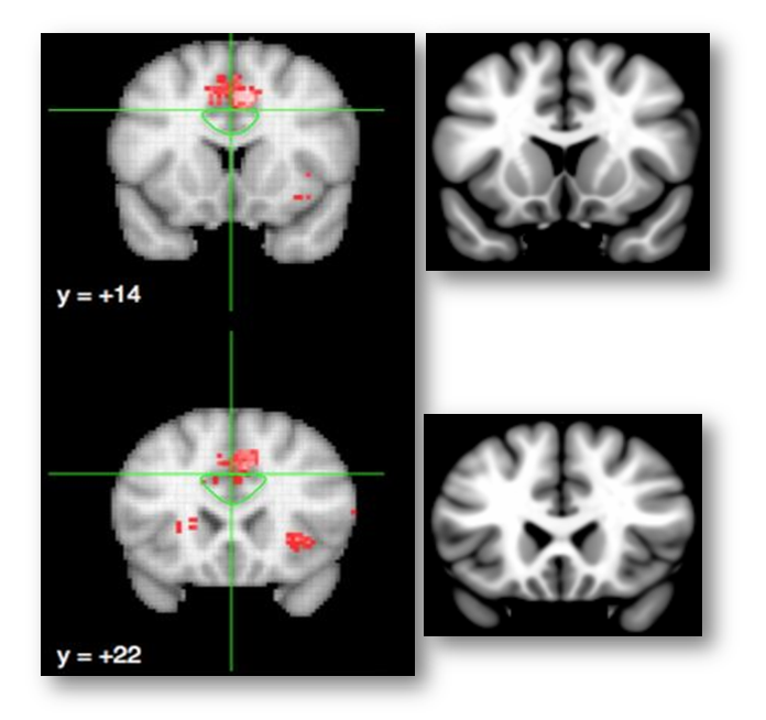
Lieberman and Eisenberger fail to account for variation in cingulate anatomy, revisited
If you accept the historical definition of dACC/MCC described above—dACC/MCC = areas 24′, 32′, and 33’—then the real problem comes from the fact that there are marked individual differences in the gross anatomy of the rostral cingulate, differences that often push area 32′ into the gray matter overlying the cingulate sulcus.
As my colleagues and I noted in a 2011 review (https://tinyurl.com/hvld5yd cited by Lieberman and Eisenberger):
“there is considerable variability in the paracingulate sulcus (PCgS), a tertiary sulcus that is present in about one-half of the population and more prominent in the left hemisphere (see the following figure, part a). The presence of this sulcus exerts a strong impact on the layout and relative volume of the architectonic areas comprising [dACC/MCC]…In particular, area 32’, which is otherwise found in the depths of the cingulate sulcus (CgS), expands to occupy the crown of the external cingulate gyrus (ECgG; the ‘superior’ or ‘paracingulate’ gyrus)…[In short] the size and spatially normalized location of the [dACC/MCC] can vary substantially across individuals…Accounting for such individual differences may permit a clearer separation of intermingled affective, nociceptive and cognitive processes [in this region]”

This is not a rare or isolated effect. As shown above, Brodmann himself depicts area 32 as lying between the cingulate (orange arrows) and paracingulate sulci (red arrows).
Likewise, Vogt and colleagues emphasize that,
“Area 24c is located on the dorsal bank of the cingulate gyrus and the ventral bank of the superior cingulate gyrus [often termed the ‘paracingulate’ gyrus] and it extends rostral to the genu but not into subgenual ACC…Area 32 is located mainly on the [paracingulate gyrus]…”
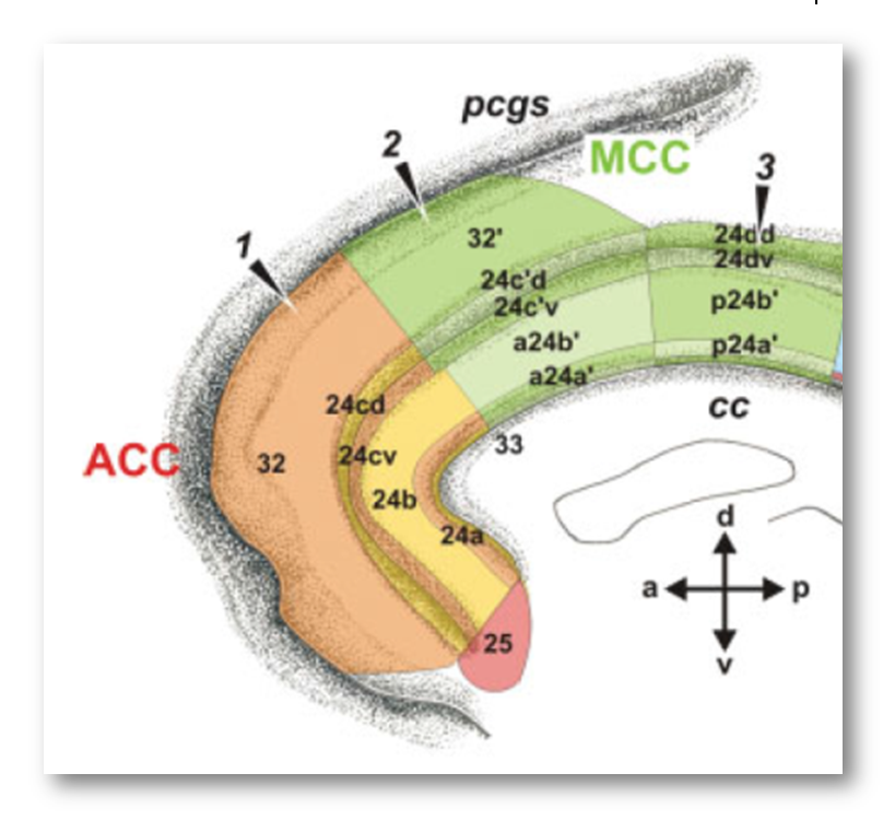
From this perspective, Lieberman and Eisenberger’s blue dACC/MCC blob, which stops at Colin’s cingulate sulcus, probably omits a substantial portion of area 32’ for many of the individual brains that underlie their meta-analyses. This is particularly problematic because cognitive neuroscientists generally regard area 32’ as the cingulate region most closely involved in cognitive conflict and control (https://tinyurl.com/p8krylv).
Lieberman and Eisenberger acknowledge these kinds of concerns in the Supplement to their report:
“There is substantial sulcal variability within the dACC….The critical question, then, is whether effects we have designated as outside the dACC…might be in the dACC after all.”
But quickly dismiss them:
“There is no way to definitively rule out this possibility in the current study. Neurosynth doesn’t have coding for individual participant morphology. Moreover, almost no fMRI studies account for these individual differences. The vast majority of fMRI studies overlook most individual differences in neuroanatomy and depend on the probabilistic neuroanatomy averaged across a group of participants and then on standard atlases that typically don’t take these individual differences into account.”
This is partially true—insofar as only a handful of fMRI studies have explicitly modeled individual differences in dACC/MCC gross anatomy (e.g. https://tinyurl.com/nsdjo3n and https://tinyurl.com/nch92eo)—but it is also deeply misleading.
In fact, as we have just seen there are at least two simple, concrete ways to begin to address variation in cingulate anatomy:
Sound like too much work? In fact, the ROI can be defined automatically using freely available probabilistic atlas labels. For example, the Harvard-Oxford atlas (https://tinyurl.com/h3m8b8w), which is included with many imaging software packages, is based on the overlap of labels generated from high-resolution anatomical scans obtained from 37 men and women of varying ages.
Applying these simple recommendations yields a very different, rather boring conclusion
Let’s see what happens when you use a probabilistic template, a probabilistic atlas, and include all of the dACC/MCC. Here is the reverse inference map reported by Lieberman and Eisenberger for studies of ‘conflict.’
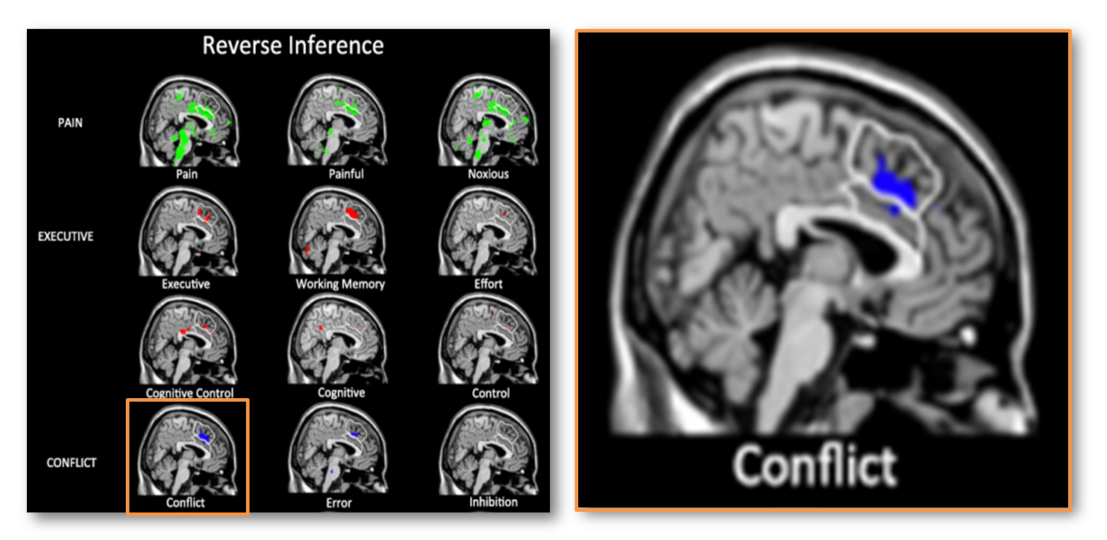
The white traces indicate their dACC/MCC (lower region) and the SMA/pre-SMA (upper region) ROI’s overlaid on Colin’s brain. Uh, oh! It looks like most of the ‘conflict’ cluster (in blue) lies in the SMA/pre-SMA (upper white region).
Here is the same map re-created using Neurosynth and overlaid on the MNI152 brain at FDR q < .01 (MNI coordinates = 7/26/35). Now it looks like the cluster (in red; colors are arbitrary) is squarely centered on the cingulate sulcus.
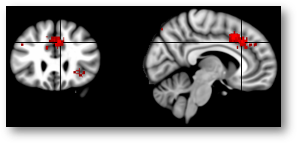
Here’s another view of the same map thresholded at Z > 7.0 to emphasize local maxima. The upper 3 panels are centered at MNI coordinates 7/26/35, which corresponds to a 46% probability of lying in the paracingulate gyrus and a 36% probability of lying in the anterior division of the cingulate gyrus in the Harvard-Oxford probabilistic atlas. The lower panel is centered at 7/15/41 (52% paracingulate, 28% anterior cingulate gyrus, and 1% supplementary motor cortex). The Harvard-Oxford atlas indicates that there is a very low probability that these maxima lie in the superior frontal gyrus or supplementary motor cortex, contrary to a SMA/pre-SMA label. Inspection of the coronal images (on the far left side of the figure) indicates that the local maxima lie in the dorsal bank of the cingulate sulcus, which is consistent with the current consensus about the role of area 32’ in monitoring cognitive conflict. My intuition is that Brodmann, Vogt, and most other investigators would label these maxima as dACC/MCC. In contrast, if we project these two points onto the sagittal plane (x=3) used in Lieberman and Eisenberger’s report and display them on Colin’s brain (right-most panels), we would draw the erroneous conclusion that they fall in the overlying SMA/pre-SMA.
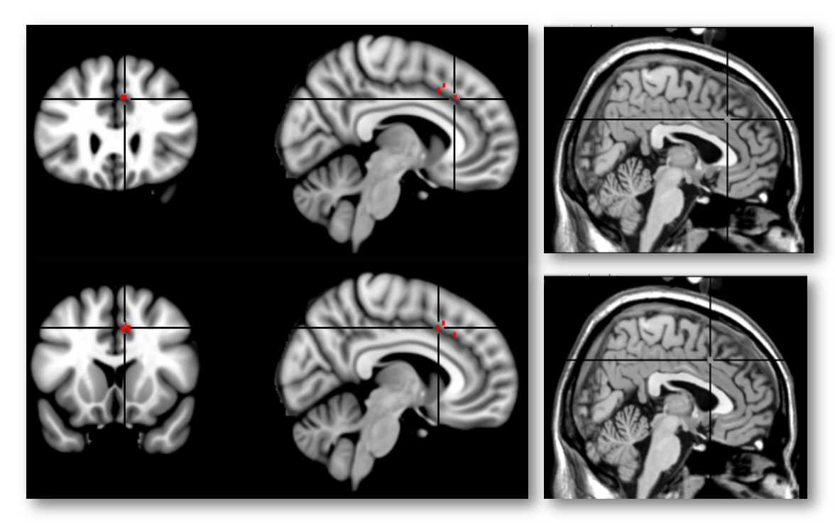
Importantly, this conclusion is not specific to ‘conflict.’ A number of other, closely related reverse-inference maps in Neurosynth show similar effects, including ‘Control’ (8/8/42; 44% cingulate gyrus anterior division, 20% paracingulate, 11% supplementary motor), ‘Errors’ (4/26/36; 63% paracingulate and 37% cingulate), ‘Stop Signal’ (6/32/34; 73% paracingulate, 9% cingulate gyrus anterior division, and 1% superior frontal gyrus), and ‘Stroop’ (6/22/38; 55% paracingulate gyrus and 27% cingulate gyrus anterior division).
In short, when probabilistic methods and longstanding definitions of the dACC/MCC are employed, we arrive at the much less extraordinary claim that tasks that demand top-down control engage the dACC/MCC.
Conclusions
So what does all of this neuroanatomy mean for Lieberman and Eisenberger’s three key claims and for past and future research more generally?
Read the guest blog, detailing recent work by our group to decipher the brain circuitry underlying extreme anxiety early in development, at EmotionNews (http://emotionnews.org/amgydala).
The week-long classroom saw students learn first-hand from subject matter experts about such topics as the function of brain neurons, the four lobes of the human brain, the effects of drugs or alcohol on an individual’s neurotransmitters and how the brain and spinal cord work together to control emotions and physical well-being.
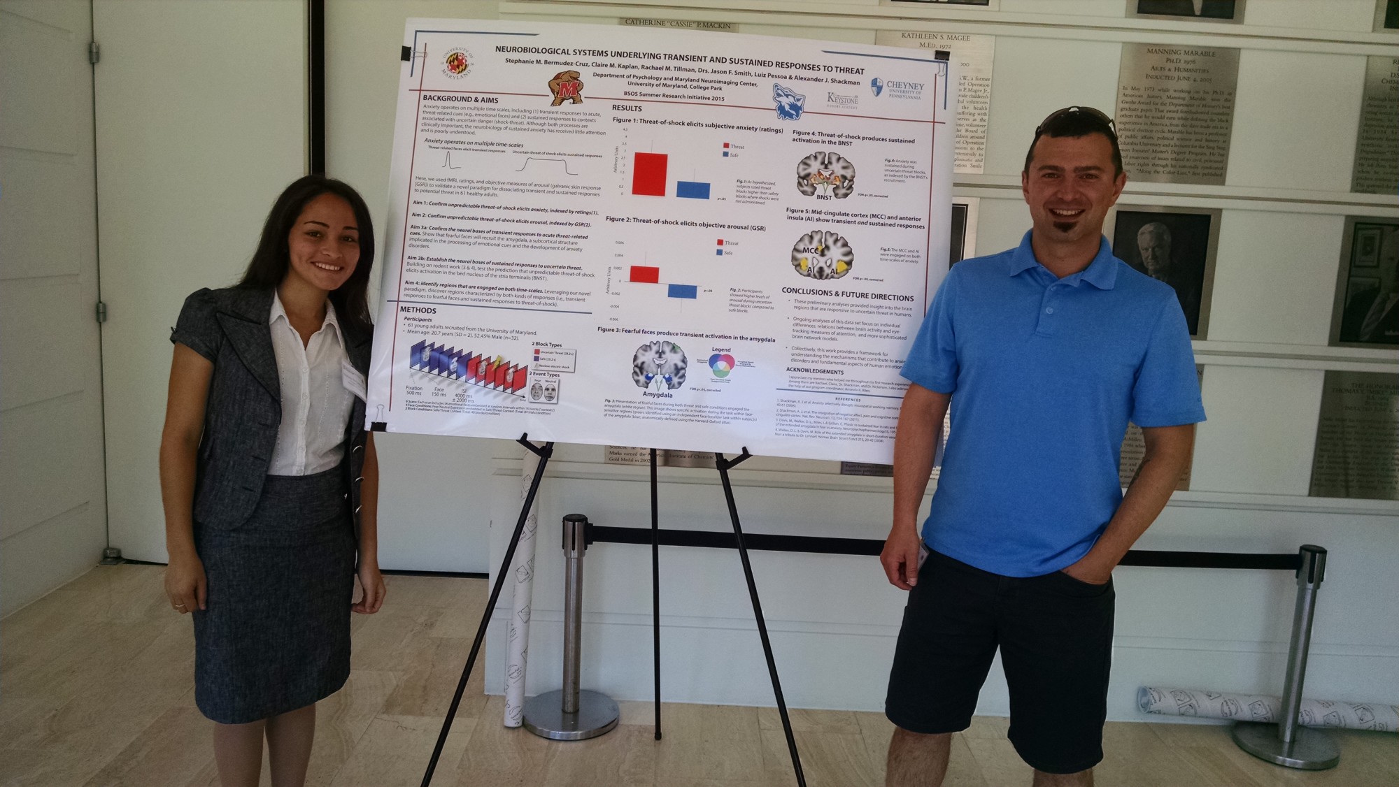
Dr. Shackman was recently elected to the board of the Neuroscience and Cognitive Science (NACS) program at UMD. To learn more, please visit the NACS website http://www.nacs.umd.edu/.
Data collection for the lab’s second fMRI study at the University of Maryland is complete. This collaborative project is focused on identifying the distributed neural circuitry underlying pervasive states of anxiety elicited by uncertain threat, a key feature of extreme dispositional anxiety and the anxiety disorders. Anxiety disorders are a leading source of human suffering and a tremendous burden on public health. These disorders, which first emerge early in life, are common, debilitating, and contribute to the development of co-morbid depression and substance abuse. Existing treatments are inconsistently effective or associated with significant adverse effects, underscoring the importance of developing a deeper understanding of the neural systems involved in sustained anxiety. This work promises to enhance our understanding of how emotional traits, like neuroticism, modulate risk, facilitate the discovery of novel biomarkers, and set the stage for developing improved interventions. From a basic psychological science perspective, our research begins to address fundamental questions about the origins of personality and temperament. Material support for this study came from the University of Maryland. Key collaborators include Luiz Pessoa (UMCP) and Matthias Gamer (University of Würzburg).
Congratulations to Jennifer Weinstein! She has recently been recognized as one of three psychology undergraduate students named among UMD’s Undergraduate Researchers of the Year for 2015. This honor includes a $1000 award, a commemorative plaque, and recognition during the opening ceremonies for Undergraduate Research Day, held from 1-4 p.m. on Wednesday, April 29 in the Grand Ballroom of the Adele H. Stamp Student Union. As a student honoree, she will be given the opportunity to present a poster on her research and accomplishments.
For more information, see:
http://go.umd.edu/UGresearchers15
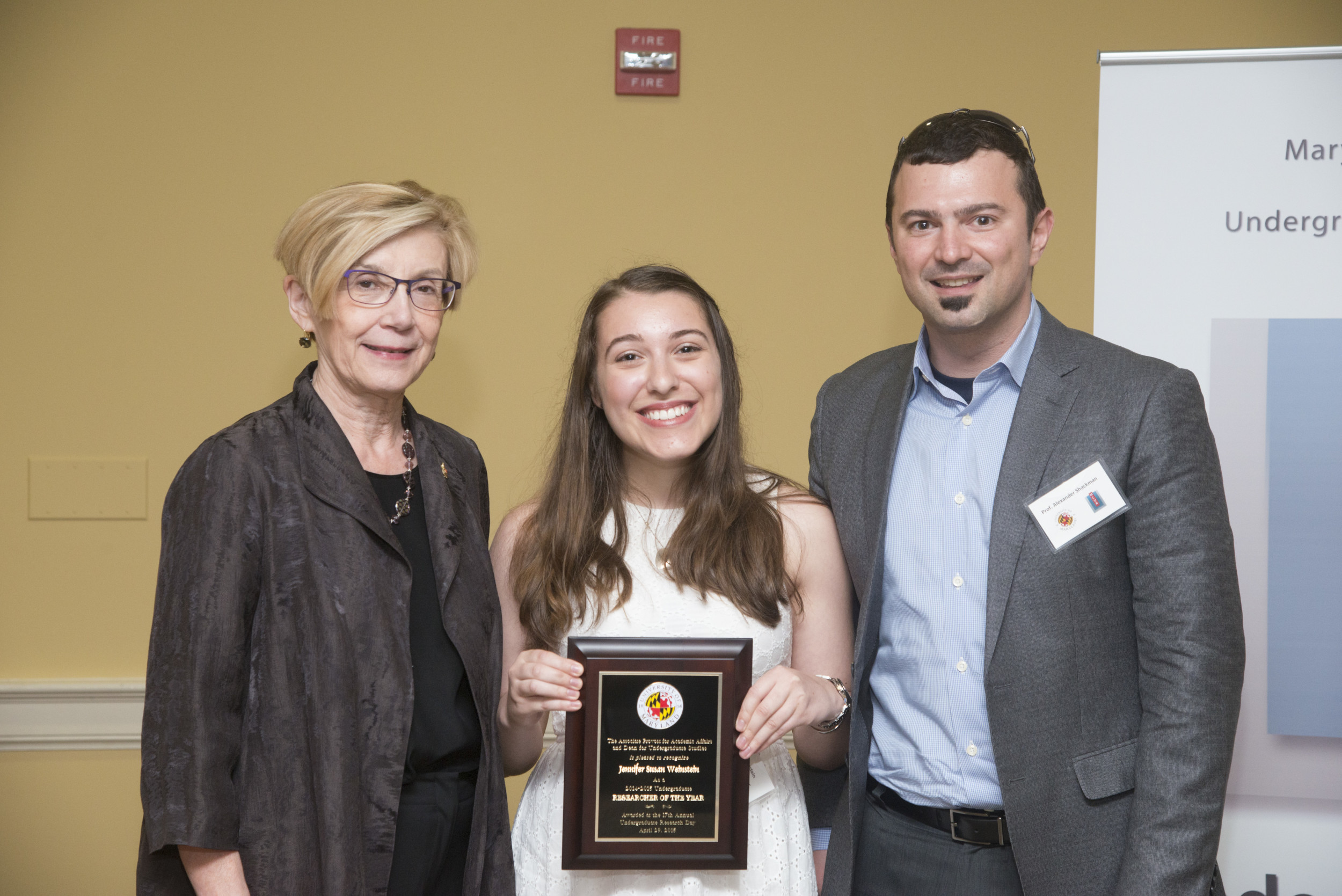
Congratulations to second year graduate student, Rachael Tillman. Rachael’s National Science Foundation Graduate Research Fellowship was highlighted for an honorable mention. Each year, ~14,000 applications are submitted by top students at research-intensive graduate programs around the country. Approximately 2,000 applications (14%) are awarded and another 2,000 receive honorable mentions.
Congratulations to Melissa Stockbridge, Rachael Tillman, and Claire Kaplan, who all successfully defended their masters project proposals. The three projects represent exciting new collaborations with Luiz Pessoa (Maryland), Andy de Los Reyes (Maryland), Talma Hendler (Tel Aviv), Leah Somerville (Harvard), and John Curtin (Wisconsin).
We’re very pleased to announce the completion of a special issue of Frontiers in Human Neuroscience devoted to The Neurobiology of Emotion-Cognition interactions.
The special issue was edited by Talma Hendler (Tel Aviv), Hadas Okon-Singer (Haifa), Luiz Pessoa (Maryland) and Dr. Shackman and features 35 reports and reviews from leading investigators in North American, Israel, and Europe—more than 100 authors in all. This work encompasses a broad spectrum of populations and showcases a wide variety of exciting new paradigms, measures, analytic strategies, and conceptual approaches.
This work appears to be making a splash—already, the 35 contributions to this Special Topic have been viewed on the Frontiers website ~90,000 times, shared or posted to social media networks >16,000 times, downloaded >13,000 times, and cited ~90 times.
For more information
Take a look at the special issue: http://journal.frontiersin.org/ResearchTopic/909
Read our Introduction to the special issue: http://journal.frontiersin.org/Journal/10.3389/fnhum.2014.01051/full
Read our commentary on The Neurobiology of Emotion-Cognition Interactions: Fundamental Questions and Strategies for Future Research: http://journal.frontiersin.org/Journal/10.3389/fnhum.2015.00058/abstract
For more information, see
Data collection for the lab’s first fMRI study conducted entirely at the University of Maryland is complete. This collaborative project is focused on identifying the neural circuitry shared by anxiety, pain, and executive cognition with a particular focus on circuits centered on the midcingulate cortex (MCC) and anterior insula (AI). In effect, the study is designed to address some of the most important challenges highlighted in Shackman et al, Nature Reviews Neuroscience, 2011. Key support for this study came from Medoc Advanced Medical Systems, which provided the thermal stimulator for delivering thermal stimulation, and the College of Behavioral & Social Sciences.
Recent results from the lab’s collaboration with Christine Larson and Danny Stout (University of Wisconsin-Milwaukee) indicate that stable individual differences in the propensity to worry, a key maladaptive feature of many anxiety disorders, reflect inefficient filtering of anxiety-related distracters from working memory, the ‘blackboard of the mind.’ The results are consistent with the idea that, once anxiety-related information gains access to this mental blackboard, it’s poised to drive future thoughts, feelings, and actions in a manner that promotes sustained distress and arousal.
D.M. Stout, A.J. Shackman, J. S. Johnson & C.L. Larson. (in press). Emotion.
Drs. Alex Shackman (PI), Luiz Pessoa, and Dave Seminowicz (Universa basic ity of Maryland, Baltimore) were awarded a Level II Dean’s Research Initiative grant from the College of Behavioral & Social Sciences. The award will support collaborative research aimed at understanding the neural circuitry shared by anxiety, pain, and executive cognition. This line of research promises to clarify the emotional and cognitive symptoms characteristic of some pain disorders. It will also set the stage for work aimed at understanding the neural circuitry supporting the inhibited, avoidant profile of choices and behaviors characteristic of many individuals with extreme anxiety.
For more information, see these recent reviews:
http://shackmanlab.org/cavanagh-j-f-shackman-a-j/#more-805
Thanks to the heroic efforts of a number of research assistants in the lab, we have completed data collection for our first large-N experience sampling study. Elevated levels of neuroticism and dispositional anxiety confer liability for the development of anxiety disorders as well as co-morbid depression and substance abuse. Here, we focused on a demographically-diverse sample of individuals selected to have a broad range of this anxious phenotype. Self-reported mood, behavior, activity, whereabouts, stress exposure, and social engagement were intensively sampled (10 surveys/day) for a week, promising an unprecedented glimpse into the daily experience of individuals at risk for developing anxiety-related psychiatric disorders. In particular, this study affords an opportunity to clarify the nature of social and behavioral inhibition “in the wild” and set the stage for linking “deep phenotype” measures to brain activity and connectivity, indexed using functional MRI.
Wonderful time at the SAS meeting catching up on the latest affective neuroscience and seeing old friends. Wonderful presentations by Tony Rangel (with an amazing introduction from Kevin Ochsner), Helen Mayberg, Richied Davidson, and Huda Akil.
Our collaborative project with Jim Cavanagh (University of New Mexico) was accepted for publication at the Journal of Physiology, Paris as part of a Special Issue focused on “Neural circuits for the adaptive control of behaviour,” edited by Jerome Sallet, Sebastien Bouret, Mark Laubach, and Dan Shulz. In the paper, Jim and I provide meta-analytic evidence that scalp-recorded control signals thought to be generated in the mid-cingulate cortex (MCC), such as the error-related negativity and N2, are consistently enhanced in dispositionally anxious individuals; and predict a more cautious, avoidant, and inhibited style of behavior following errors and punishment. The meta-analysis incorporated studies of more than 2,000 research subjects, enhancing our confidence in these results. These observations reinforce the idea that a circuit centered on the MCC plays a central role in regulating behavior in the face of uncertainty about instrumental actions and their potentially aversive outcomes and set the stage for future research aimed at delineating the neural mechanisms underlying the maladaptive profile of choices and actions characteristic of many individuals with elevated anxiety.
Cavanagh, J. F. & Shackman, A. J. (in press). Frontal midline theta reflects anxiety and cognitive control: Meta-analytic evidence. Journal of Physiology, Paris.”
Our resting-state fMRI paper was just accepted for publication at Molecular Psychiatry (2012 Impact Factor 14.897; #1/135 Psychiatry). In the report, we provide new evidence that extreme anxiety early in life is associated with aberrant functional integration between the central nucleus of the amygdala and prefrontal cortex. This pattern was discernible in lightly anesthetized young rhesus monkeys as well as quietly resting children with diagnosed anxiety disorders, indicating an evolutionarily conserved circuit, and opening the door to future mechanistic research. In the large sample of young non-human primates (n = 89), reduced functional connectivity was associated with elevated metabolic activity in the amygdala and chronically heightened anxiety, behavioral inhibition, and cortisol outside of the scanner. From a translational perspective, these findings provide a novel neurobiological framework for conceptualizing extreme anxiety and set the stage for the development of novel therapeutic strategies. From the vantage point of basic psychological science, they suggest that core features of our temperament and personality are embodied in the spontaneous, on-going activity of the brain.
The ADAA rolled out an exceptionally useful early-career professional development program. Excellent presentation by Charlie Nemeroff on grantsmanship and plenty of one-on-time to talk about funding strategies, lab management/administration, and other key topics with Ned Kalin, Kerry Ressler, and a fabulous group of young scientists and clinicians.
A very warm welcome to our three new graduate students: Claire Kaplan (Weinberger lab at Hopkins), Rachael Tillman (McPartland lab at the Yale Child Study Center), and Melissa Stockbridge (Newman lab, UMCP). Claire and Rachael will be starting in the clinical Ph.D. program in the Fall of 2014. Melissa is currently a doctoral student in the Department of Hearing & Speech Sciences and will be co-supervised by Dr. Shackman during the 2014-15 academic year.
We (http://shackmanlab.org) are looking for several undergraduate research assistants (RA’s) to assist with on-going projects focused on the neural substrates of anxiety. PHONES: One project uses mobile phone technology to assess anxious mood and behavior in the field. For the mobile phone study, we need RA’s available M/W 10:45am-1:15pm &/or T/R afternoon 1:45-5:15pm. fMRI: The other project uses fMRI to quantify anxiety-related brain activity and connectivity. For the fMRI study, we need RA’s available T/R 1-6pm &/or W 9am-4pm. Contact Dr. Shackman directly if you are interested (shackman@umd.edu).
Oxford University Press has agreed to publish a new edition of The Nature of Emotion: Fundamental Questions, to be co-edited by Richie Davidson, Drew Fox, Regina Lapate, and Alex Shackman. The first edition, edited by Davidson and Paul Ekman and published in 1995, was a landmark in the study of emotion (http://www.amazon.com/The-Nature-Emotion-Fundamental-Questions/dp/0195089448). We’re immensely excited about the new edition, which will retain the unique question and short answer format of its predecessor, while delving more deeply into the neurobiology of emotion.
Dr. Shackman will be chairing symposia focused on the neural substrates of extreme anxiety at the annual meetings of the Anxiety and Depression Association of America as well the Society of Biological Psychiatry. Other participants include Luiz Pessoa, Talma Hendler, Danny Pine, Leah Somerville, Dylan Gee, Drew Fox, and Ned Kalin. Join us for what promises to be two very exciting sets of talks.
Dr. Alex Shackman has been selected to participate in the Anxiety and Depression Association of America’s Career Development Leadership Program. This is a highly competitive program and Dr. Shackman was selected from an extraordinarily accomplished application pool. Congratulations to Dr. Shackman!
Assistant Professor Alex Shackman has been appointed to the editorial boards of the APA journal Emotion and the Psychonomic Society’s journal Cognitive, Affective, and Behavioral Neuroscience
We’re pleased to welcome Dr. Jason Smith, our new imaging data analyst, to the ATNL family. Jason brings a decade’s worth of experience with functional MRI processing and analysis, most recently in the Brain Imaging and Modeling Section, NIDCD.
My lab is looking to hire a programmer / IT staff person. We’re open to this being a position filled by an IT savvy post-bac prior to grad school, a grad student/postdoc looking to switch off the academic track, someone with a traditional IT background, or anything in between. Please have a very low threshold for contacting me to learn more about the position or suggest someone you know.